Automatic Initialization Active Contour Model for the Segmentation of the Chest Wall on Chest CT
- Affiliations
-
- 1Department of Radiological Science, College of Health Sciences, Catholic University of Pusan, Busan, Korea. cszzim@cup.ac.kr
- KMID: 2166564
- DOI: http://doi.org/10.4258/hir.2010.16.1.36
Abstract
OBJECTIVES
Snake or active contours are extensively used in computer vision and medical image processing applications, and particularly to locate object boundaries. Yet problems associated with initialization and the poor convergence to boundary concavities have limited their utility. The new method of external force for active contours, which is called gradient vector flow (GVF), was recently introduced to address the problems. METHODS: This paper presents an automatic initialization value of the snake algorithm for the segmentation of the chest wall. Snake algorithms are required to have manually drawn initial contours, so this needs automatic initialization. In this paper, our proposed algorithm is the mean shape for automatic initialization in the GVF. RESULTS: The GVF is calculated as a diffusion of the gradient vectors of a gray-level or binary edge map derived from the medical images. Finally, the mean shape coordinates are used to automatic initialize thepoint of the snake. The proposed algorithm is composed of three phases: the landmark phase, the procrustes shape distance metric phase and aligning a set of shapes phase. The experiments showed the good performance of our algorithm in segmenting the chest wall by chest computed tomography. CONCLUSIONS: An error analysis for the active contours results on simulated test medical images is also presented. We showed that GVF has a large capture range and it is able to move a snake into boundary concavities. Therefore, the suggested algorithm is better than the traditional potential forces of image segmentation.
Keyword
Figure
-
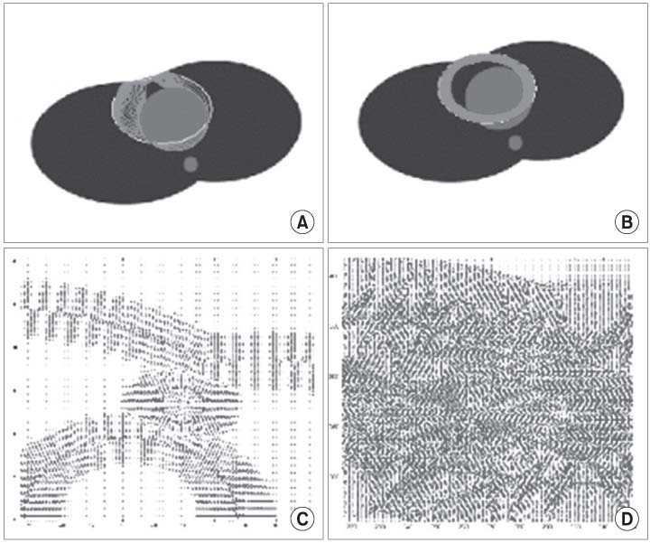
Figure 1 Vector field changes of chest wall model in the µ variation. (A) µ = 0.02, iter. = 60. (B) µ = 0.2, iter. = 60. (C) µ = 0.02, iter. = 60. (D) µ = 0.2, iter. = 60. (A, B) shows initialization and convergence to boundary concavity.
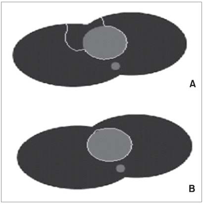
Figure 2 Vector field changes of chest wall model in the α variation. (A) α = 0.001, µ = 0.2, iter. = 60. (B) α = 0.2, µ = 0.2, iter. = 60. White circle in the (B) represent final segmentation result.
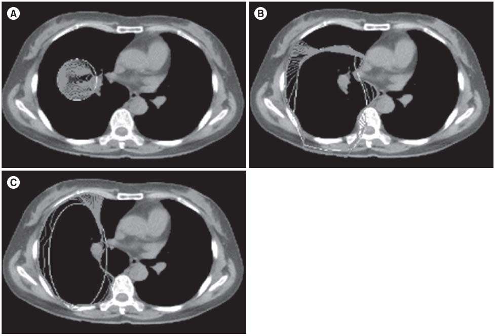
Figure 3 Result in the initial curve value (deformation in progress, iter. = 500). (A) manual initialization, (B) ellipse, automatic initialization, (C) ellipse, automatic initialization.

Figure 4 Steps of the mean shape.
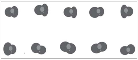
Figure 5 Chest wall image of pseudo model for training set (chest deformation).
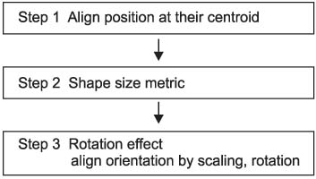
Figure 6 Steps of the procrustes shape.
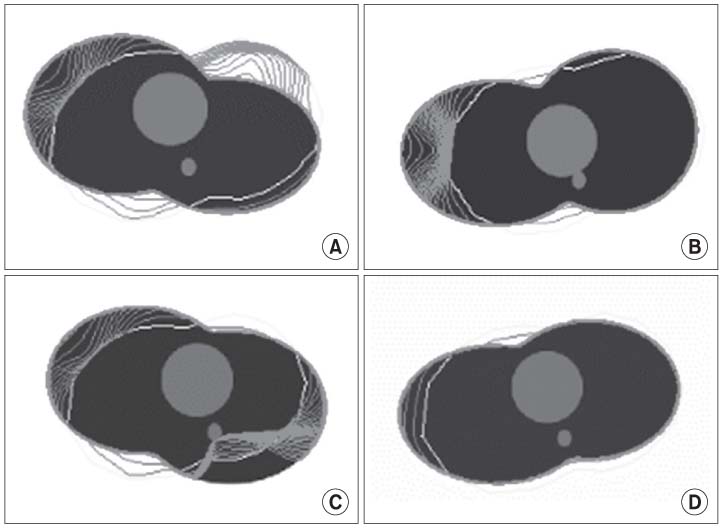
Figure 7 Vector field changes of procrustes shape in the auto initialization (pseudo chest wall image). (A, B) µ = 0.2, iter. = 60, (C, D) µ = 0.2, iter. = 60. (A-D) shows initialization and convergence to boundary concavity.
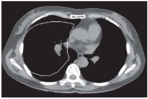
Figure 8 Auto initialization computing the procrustes shape (chest wall image).

Figure 9 Snake convergence with the auto initialization.

Figure 10 Boundary concavity of chest wall with the auto initialization.

Figure 11 Total calculation time of image segmentation in the auto initialization and manual.
Reference
-
1. Nappi J, Dachman AH, MacEneaney P, Yoshida H. Automated knowledge-guided segmentation of colonic walls computerized detection of polyps in CT colonography. J Comput Assist Tomogr. 2002. 26:493–504.2. Henkel RD. Segmentation in scale space. Computer analysis of images and patterns (Lecture Notes in Computer Science. Vol. 970). 1995. Berlin: Springer-Verlag;41–48.
Article3. Barish MA, Rocha TC. Multislice CT colonography: current status and limitations. Radiol Clin North Am. 2005. 43:1049–1062.
Article4. Gao H, Siu WC, Hou CH. Improved techniques for automatic image segmentation. IEEE Trans Circuits Syst Video Technol. 2001. 11:1273–1280.
Article5. Ballard DH, Brown CM. Computer vision. 1982. Englewood Cliffs: Prentice-Hall;116–123.6. Kass M, Witkin A, Terzopoulos D. Snakes: active contour models. Int J Comput Vis. 1988. 1:321–331.
Article7. Xu C, Prince JL. Generalized gradient vector flow external forces for active contours. Signal Process. 1998. 71:131–139.
Article8. Ma L, Zhu L. Liver contour extraction using snake and initial boundary auto-generation. 2008. In : Proceedings of the 2nd International Conference on Bioinformatics and Biomedical Engineering; Shanghai, China. 2669–2672.
Article9. Mala K, Sadasivam V. Automatic segmentation and classification of diffused liver diseases using wavelet based texture analysis andneural network. 2005. In : Proceedings of IEEE Indicon 2005 Conference; 2005 Dec 11-13; Chennai, IN. Los Alamitos (CA): IEEE Computer Society;216–219.10. McInerney T, Terzopoulos D. Deformable models in medical image analysis: a survey. Med Image Anal. 1996. 1:91–108.
Article11. Lobregt S, Viergever MA. A discrete dynamic contour model. IEEE Trans Med Imaging. 1995. 14:12–24.
Article12. Hwang YH. Locating chest boundary in sequential images by snakes [dissertation]. 1997. Seoul: Myongji University.13. Li C, Liu J, Fox M. In : Schmid C, Soatto S, Tomasi C, editors. Segmentation of edge preserving gradient vector flow: an approach toward automatically initializing and splitting of snakes. 2005. Proceedings of the 2005 IEEE Computer Society Conference on Computer Vision and Pattern Recognition (CVPR'05); 2005 June 20-25; San Diego, CA. Los Alamitos (CA): IEEE Computer Society.
Article14. Cootes TF, Taylor CJ, Cooper DH, Graham J. Active shape models: their training and application. Comput Vis Image Underst. 1995. 61:38–59.15. Gower JC, Dijksterhuis GB. Procrustes problems. 2004. New York: Oxford University Press;29–83.16. Biniwale RM, Weinstein S, Boulanger SC, Glick P. eMedicine: Chest wall deformities [Internet]. 1994-2010. [cited 2009 Jan 1]. Medscape;Available from http://emedicine.medscape.com/article/906078-overview.17. Park JS, Hwang SB, Chung MS, Shin DS, Park HS, Lee YS, et al. Three dimensional automatic surface reconstruction software. J Korean Soc Med Inform. 2007. 13:385–392.
Article
- Full Text Links
-
- Actions
-
Cited
- CITED
-
- Close
- Share
-
- Similar articles
-
- Evaluation of Semi-automatic Segmentation Methods for Persistent Ground Glass Nodules on Thin-Section CT Scans
- Multiple hamartomas(mesenchymomas) of the unilateral chest wall in infancy: CT findings
- Segmentation of Hippocampus in Human Brain MRI Using Watersnakes
- Multi-Detector CT Findings of Palpable Chest Wall Masses in Children: A Pictorial Essay
- The Clinical Feature and Pressure Threshold in a Chest Wall Syndrome
