Cancer Res Treat.
2010 Jun;42(2):115-117.
A Case of Pure Red Cell Aplasia Associated with Angioimmunoblastic T-cell Lymphoma
- Affiliations
-
- 1Department of Internal Medicine, Hanyang University College of Medicine, Seoul, Korea. jhcmd@hanyang.ac.kr
- 2Department of Pathology, Hanyang University College of Medicine, Seoul, Korea.
- 3Department of Laboratory Medicine, Hanyang University College of Medicine, Seoul, Korea.
Abstract
- Pure red cell aplasia is a bone marrow failure characterized by a progressive normocytic anemia and reticulocytopenia without leucopenia and thrombocytopenia. It is associated with various hematologic diseases. However, pure red cell aplasia with angioimmunoblastic T cell lymphoma has rarely been reported. Here we describe a 43-year-old woman with pure red cell aplasia associated with angioimmunoblastic T-cell lymphoma. She had severe anemia (hemoglobin 6.9 g/dL) and a low reticulocyte count (0.2%). Direct and indirect Coombs' tests were positive. A CT scan of the abdomen revealed marked hepatosplenomegaly and small multiple lymphadenopathies. A bone marrow biopsy revealed focal infiltration of abnormal lymphoid cells and absence of red cell precursors. Splenic biopsy was compatible with angioimmunoblastic T-cell lymphoma. Ultimately, diagnosis of pure red cell aplasia associated with angioimmunoblastic T-cell lymphoma was made. After initiating CHOP therapy, the patient achieved complete remission, which was accompanied, shortly thereafter, by a rise in hemoglobin levels which finally returned to normal.
Keyword
MeSH Terms
Figure
-
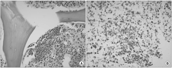
Fig. 1 (A) Bone marrow biopsy shows focal infiltration of abnormal lymphoid cells and the absence of red cell precursors (H&E, ×400). (B) Splenic biopsy shows diffuse infiltration of atypical lymphoid cells which are positive for CD3 (CD3, ×400).
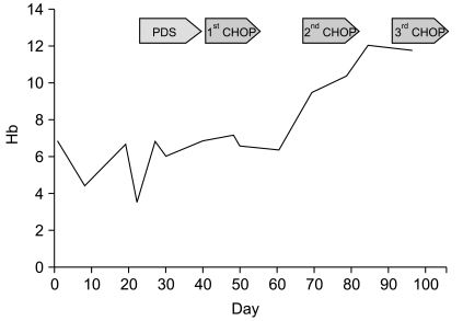
Fig. 2 Change of hemoglobin levels during treatment.
Reference
-
1. Djaldetti M, Blay A, Bergman M, Salman H, Bessler H. Pure red cell aplasia--a rare disease with multiple causes. Biomed Pharmacother. 2003; 57:326–332. PMID: 14568226.
Article2. Lynch JW, Elfenbein GJ, Noyes WD, Braylan RC, Gross MA, Weiner RS. Pure red cell aplasia associated with angioimmunoblastic lymphadenopathy with dysproteinemia. Am J Hematol. 1994; 46:72–78. PMID: 8172198.
Article3. Tsujimura H, Sakai C, Takagi T. Pure red cell aplasia complicated by angioimmunoblastic T-cell lymphoma: humoral factor plays a main role in the inhibition of erythropoiesis from CD34 (+) progenitor cells. Am J Hematol. 1999; 62:259–260. PMID: 10589086.4. Higuchi T, Mori H, Niikura H, Omine M. Hypocomplementemia and hematological abnormalities in immunoblastic lymphadenopathy and immunoblastic lymphadenopathy-like T cell lymphoma. Acta Haematol. 1996; 96:68–72. PMID: 8701703.
Article5. Alberti E, Aldovini D, Mazzon C, Dal Rì P, Rubertelli M. Angioimmunoblastic Lymphadenopathy with Dysproteinemia. Report of a case with pure red cell aplasia. Haematologica. 1982; 67:919–925. PMID: 6819197.
- Full Text Links
-
- Actions
-
Cited
- CITED
-
- Close
- Share
-
- Similar articles
-
- Pure Red Cell Aplasia Caused by Acute Hepatitis A
- Pure Red Cell Aplasia Associated with Diphenylhydantoin: Case Report
- Drug-induced hepatitis complicated by pure red cell aplasia and autoimmune hemolytic anemia: a case report
- Pure Red Cell Aplasia in Pregnancy
- A case of AILD associated with pure red cell aplasia
