Correlation of Narrow Band Imaging with Magnifying Colonoscopy and Histology in Colorectal Tumors
- Affiliations
-
- 1Department of Internal Medicine, Soonchunhyang University Bucheon Hospital, Soonchunhyang University College of Medicine, Bucheon, Korea. mslee@schmc.ac.kr
- 2Department of Pathology, Soonchunhyang University Bucheon Hospital, Soonchunhyang University College of Medicine, Bucheon, Korea.
Abstract
- BACKGROUND/AIMS
Narrow band imaging (NBI) is a new technique that uses optical filters for imaging of mucosal morphology. The aim of this study was to correlate findings of NBI with magnifying colonoscopy and histology for prediction of neoplastic colorectal lesion.
METHODS
Between September 2005 and December 2007, 107 colon polyps from 68 patients were detected by conventional colonoscopy and subsequently evaluated by NBI with magnifying colonoscopy and analyzed for a pit pattern and a capillary pattern. More analysis was done regarding thickness and irregularity of capillary features.
RESULTS
Pit pattern with NBI magnification to discriminate between neoplastic and non-neoplastic lesions had a sensitivity of 88.9% and a specificity of 87.5%; capillary pattern yielded test performance characteristics of 91.9% and 87.5%. In respect of capillary thickness, invisible capillaries were found significantly more often in hyperplastic lesions. All thick capillaries were found in neoplastic polyps, and found significantly more often in carcinomas with submucosal massive invasion (sm-m) (p<0.01). In respect of capillary irregularity, invisible capillaries were found significantly more often in hyperplasic lesions, and severely irregular capillaries were found significantly more often in sm-m lesions (p<0.01).
CONCLUSIONS
Observation of capillary thickness and irregularity by NBI magnification is useful for correlating histological grade with carcinoma, especially with depth of submucosal invasion.
MeSH Terms
Figure
-
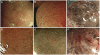
Fig. 1 Classification of capillary pattern. (A, B) Capillary pattern type I shows absence of meshed brown capillary vessel. (C, D) Capillary pattern type II shows presence of meshed brown capillary vessel, slightly thicker and loose capillary density. (E, F) Capillary pattern type III shows presence of meshed brown capillary vessel, thicker, branching, irregularity of capillary and dense capillary density.
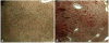
Fig. 2 Classification of microvessel thickness as determined by narrow band imaging. (A) Microvessels were classified as thin when they were thin and even thickness. (B) Microvessels were classified as thick when they were uneven thickness.
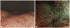
Fig. 3 Classification of microvascular features as determined by narrow band imaging. (A) Microvessels were classified regular when lesions had microvessels running smoothly between pits and an apparent regular meshwork of microvessels throughout. (B) Microvessels were classified irregular when lesions had microvessels running irregularly between pits and an apparent irregular meshwork of microvessels throughout.
Reference
-
1. Sano Y, Muto M, Tajiri H, Ohtsu A, Yoshida S. Optical/digital chromoendoscopy during colonoscopy using narrow-band imaging system. Dig Endosc. 2005; 17(Suppl 1):S43–S48.
Article2. Gono K, Obi T, Yamaguchi M, et al. Appearance of enhanced tissue features in narrow-band endoscopic imaging. J Biomed Opt. 2004; 9:568–577. PMID: 15189095.
Article3. Machida H, Sano Y, Hamamoto Y, et al. Narrow-band imaging in the diagnosis of colorectal mucosal lesions: a pilot study. Endoscopy. 2004; 36:1094–1098. PMID: 15578301.
Article4. East JE, Suzuki N, Bassett P, et al. Narrow band imaging with magnification for the characterization of small and diminutive colonic polyps: pit pattern and vascular pattern intensity. Endoscopy. 2008; 40:811–817. PMID: 18828077.
Article5. Sikka S, Ringold DA, Jonnalagadda S, Banerjee B. Comparison of white light and narrow band high definition images in predicting colon polyp histology, using standard colonoscopes without optical magnification. Endoscopy. 2008; 40:818–822. PMID: 18668472.
Article6. Bansal A, Ulusarac O, Mathur S, Sharma P. Correlation between narrow band imaging and nonneoplastic gastric pathology: a pilot feasibility trial. Gastrointest Endosc. 2008; 67:210–216. PMID: 18226682.
Article7. Yao K, Iwashita A, Tanabe H, et al. White opaque substance within superficial elevated gastric neoplasia as visualized by magnification endoscopy with narrow-band imaging: a new optical sign for differentiating between adenoma and carcinoma. Gastrointest Endosc. 2008; 68:574–580. PMID: 18656862.
Article8. Kitajima K, Fujimori T, Fujii S, et al. Correlations between lymph node metastasis and depth of submucosal invasion in submucosal invasive colorectal carcinoma: a Japanese collaborative study. J Gastroenterol. 2004; 39:534–543. PMID: 15235870.
Article9. Kudo S, Tamura S, Nakajima T, Yamano H, Kusaka H, Watanabe H. Diagnosis of colorectal tumorous lesions by magnifying endoscopy. Gastrointest Endosc. 1996; 44:8–14. PMID: 8836710.
Article10. Sano Y, Horimatsu T, Fu K, Katagiri A, Muto M, Ishikawa H. Magnifying observation of microvascular architecture of colorectal lesions using a narrow-band system. Dig Endosc. 2006; 18(Suppl 1):S44–S51.11. Tanaka S, Haruma K, Nagata S, Oka S, Chayama K. Diagnosis of invasion depth in early colorectal carcinoma by pit pattern analysis with magnifying endoscopy. Dig Endosc. 2001; 13(Suppl 1):S2–S5.
Article12. Tanaka S, Haruma K, Ito M, et al. Detailed colonoscopy for detecting early superficial carcinoma: recent developments. J Gastroenterol. 2000; 35(Suppl 12):121–125. PMID: 10779231.13. Kudo S, Hirota S, Nakajima T, et al. Colorectal tumours and pit pattern. J Clin Pathol. 1994; 47:880–885. PMID: 7962600.
Article14. Oka S, Tanaka S, Nagata S, et al. Relationship between histopathological features and type V pit pattern determined by magnifying video colonoscopy in early colorectal carcinoma. Dig Endosc. 2005; 17:117–122.15. Nagata S, Tanaka S, Haruma K, et al. Pit pattern diagnosis of early colorectal carcinoma by magnifying colonoscopy: clinical and histological implications. Int J Oncol. 2000; 16:927–934. PMID: 10762628.
Article16. Hata K, Watanabe T, Motoi T, Nagawa H. Pitfalls of pit pattern diagnosis in ulcerative colitis-associated dysplasia. Gastroenterology. 2004; 126:374–376. PMID: 14753219.
Article17. Kiesslich R, Fritsch J, Holtmann M, et al. Methylene blue-aided chromoendoscopy for the detection of intraepithelial neoplasia and colon cancer in ulcerative colitis. Gastroenterology. 2003; 124:880–888. PMID: 12671882.
Article18. Larghi A, Lecca PG, Costamagna G. High-resolution narrow band imaging endoscopy. Gut. 2008; 57:976–986. PMID: 18208902.
Article19. Inoue T, Murano M, Murano N, et al. Comparative study of conventional colonoscopy and pan-colonic narrow-band imaging system in the detection of neoplastic colonic polyps: a randomized, controlled trial. J Gastroenterol. 2008; 43:45–50. PMID: 18297435.
Article20. Tanaka S, Oka S, Hirata M, Yoshida S, Kaneko I, Chayama K. Pit pattern diagnosis for colorectal neoplasia using narrow band imaging magnification. Dig Endosc. 2006; 18(Suppl 1):S52–S56.
Article21. Su MY, Hsu CM, Ho YP, Chen PC, Lin CJ, Chiu CT. Comparative study of conventional colonoscopy, chromoendoscopy, and narrow-band imaging systems in differential diagnosis of neoplastic and nonneoplastic colonic polyps. Am J Gastroenterol. 2006; 101:2711–2716. PMID: 17227517.
Article22. Nakayoshi T, Tajiri H, Matsuda K, Kaise M, Ikegami M, Sasaki H. Magnifying endoscopy combined with narrow band imaging system for early gastric cancer: correlation of vascular pattern with histopathology (including video). Endoscopy. 2004; 36:1080–1084. PMID: 15578298.
Article23. Hirata M, Tanaka S, Oka S, et al. Magnifying endoscopy with narrow band imaging for diagnosis of colorectal tumors. Gastrointest Endosc. 2007; 65:988–995. PMID: 17324407.
Article24. Hirata M, Tanaka S, Oka S, et al. Evaluation of microvessels in colorectal tumors by narrow band imaging magnification. Gastrointest Endosc. 2007; 66:945–952. PMID: 17963882.
Article25. Rastogi A, Bansal A, Wani S, et al. Narrow-band imaging colonoscopy: a pilot feasibility study for the detection of polyps and correlation of surface patterns with polyp histologic diagnosis. Gastrointest Endosc. 2008; 67:280–286. PMID: 18155210.26. Sano Y, Horimatsu T, Fu KI, et al. Magnified observation of micro-vascular architecture using narrow band imaging (NBI) for the differential diagnosis between non-neoplastic and neoplastic colorectal lesion: a prospective study. Gastrointest Endosc. 2006; 63(5 Suppl):AB102.
Article
- Full Text Links
-
- Actions
-
Cited
- CITED
-
- Close
- Share
-
- Similar articles
-
- Computer-aided System for Predicting the Histology of Colorectal Tumors by Using Narrow-band Imaging Magnifying Colonoscopy (Gastrointest Endosc 2012;75:179-185)
- Polyp Detection, Characterization, and Management Using Narrow-Band Imaging with/without Magnification
- Clinical Usefulness of Magnifying Chromoendoscopy and Magnifying Narrow Band Imaging Endoscopy for Predicting the Submucosal Invasion of Early Colorectal Cancers
- Artificial Intelligence-Based Colorectal Polyp Histology Prediction by Using Narrow-Band Image-Magnifying Colonoscopy
- Endoscopic Assessment of Colorectal Cancer with Superficial or Deep Submucosal Invasion Using Magnifying Colonoscopy
