Clin Endosc.
2013 Sep;46(5):595-597.
Retroperitoneal Cystic Lymphangioma Diagnosed by Endoscopic Ultrasound-Guided Fine Needle Aspiration
- Affiliations
-
- 1Department of Internal Medicine, Duke University Medical Center, Durham, NC, USA. tylerpblack@gmail.com
- 2Department of Pathology, Duke University Medical Center, Durham, NC, USA.
- 3Department of Gastroenterology, Duke University Medical Center, Durham, NC, USA.
Abstract
- Retroperitoneal cystic lymphangiomas are rare tumors of the lymphatic system. These tumors usually present in childhood and are often diagnosed incidentally with imaging procedures. Although benign, they can grow to large sizes and become symptomatic due to their compressive effects. They can cause diagnostic dilemmas with other retroperitoneal cystic tumors including those arising from the liver, kidney, and pancreas. Endoscopic ultrasound (EUS) has become an invaluable tool in the assessment of cystic lesions in the region of the pancreas. This case describes a 66-year-old female who presented with 3 months of abdominal pain. Radiographic imaging was suggestive of a cystic lesion in the region of the pancreas. EUS was performed confirming a cystic lesion adjacent to the tail of the pancreas with subsequent fine needle aspiration fluid analysis consistent with a cystic lymphangioma.
Keyword
MeSH Terms
Figure
-
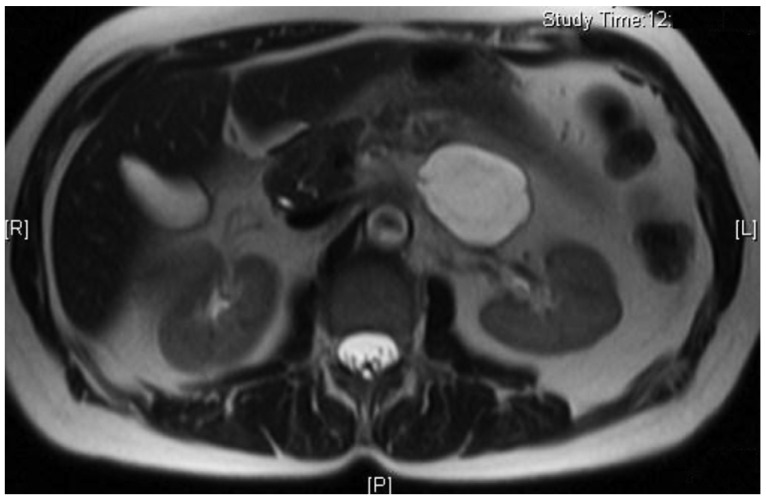
Fig. 1 Magnetic resonance imaging abdomen showing cystic lesion in region of pancreas.

Fig. 2 Endoscopic ultrasound showing cystic lesion adjacent to pancreas.
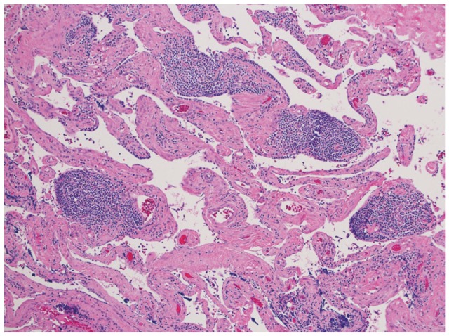
Fig. 3 H&E stain at ×100 magnification shows variably sized, dilated endothelial-lined spaces with a hypocellular, fibrovascular connective tissue stroma, and collections of lymphocytes.

Fig. 4 Immunohistochemistry for D240, a marker for lymphatic endothelium (×100).
Reference
-
1. Hayami S, Adachi Y, Ishigooka M, et al. Retroperitoneal cystic lymphangioma diagnosed by computerized tomography, magnetic resonance imaging and thin needle aspiration. Int Urol Nephrol. 1996; 28:21–26. PMID: 8738615.
Article2. Bhavsar T, Saeed-Vafa D, Harbison S, Inniss S. Retroperitoneal cystic lymphangioma in an adult: a case report and review of the literature. World J Gastrointest Pathophysiol. 2010; 1:171–176. PMID: 21607159.
Article3. Nuzzo G, Lemmo G, Marrocco-Trischitta MM, Boldrini G, Giovannini I. Retroperitoneal cystic lymphangioma. J Surg Oncol. 1996; 61:234–237. PMID: 8637214.
Article4. Shankar KR, Roche CJ, Carty HM, Turnock RR. Cystic retroperitoneal lymphangioma: treatment by image-guided percutaneous catheter drainage and sclerotherapy. Eur Radiol. 2001; 11:1021–1023. PMID: 11419147.
Article5. de Perrot M, Rostan O, Morel P, Le Coultre C. Abdominal lymphangioma in adults and children. Br J Surg. 1998; 85:395–397. PMID: 9529502.
Article6. Coe AW, Evans J, Conway J. Pancreas cystic lymphangioma diagnosed with EUS-FNA. JOP. 2012; 13:282–284. PMID: 22572132.7. Henzel JH, Pories WJ, Burget DE, Smith JL. Intra-abdominal lymphangiomata. Arch Surg. 1966; 93:304–308. PMID: 5913566.
Article8. Jathal A, Arsenescu R, Crowe G, Movva R, Shamoun DK. Diagnosis of pancreatic cystic lymphangioma with EUS-guided FNA: report of a case. Gastrointest Endosc. 2005; 61:920–922. PMID: 15933705.
Article9. Yagihashi Y, Kato K, Nagahama K, Yamamoto M, Kanamaru H. A case of laparoscopic excision of a huge retroperitoneal cystic lymphangioma. Case Rep Urol. 2011; 2011:712520. PMID: 22606623.
Article
- Full Text Links
-
- Actions
-
Cited
- CITED
-
- Close
- Share
-
- Similar articles
-
- Endoscopic Ultrasound-Guided Fine Needle Aspiration in Cystic Pancreatic Lesions
- Fine-Needle Biopsy: Should This Be the First Choice in Endoscopic Ultrasound-Guided Tissue Acquisition?
- How Can We Get the Best Results with Endoscopic Ultrasound-Guided Fine Needle Aspiration?
- Endoscopic Ultrasound-Fine Needle Aspiration versus Core Biopsy for the Diagnosis of Subepithelial Tumors
- Role of Repeated Endoscopic Ultrasound-Guided Fine Needle Aspiration for Inconclusive Initial Cytology Result
