Optical Molecular Imaging for Diagnosing Intestinal Diseases
- Affiliations
-
- 1Asan Institute for Life Sciences, Asan Medical Center, University of Ulsan College of Medicine, Seoul, Korea. sjmyung@amc.seoul.kr
- 2Department of Gastroenterology, Asan Medical Center, University of Ulsan College of Medicine, Seoul, Korea.
Abstract
- Real-time visualization of the molecular signature of cells can be achieved with advanced targeted imaging techniques using molecular probes and fluorescence endoscopy. This molecular optical imaging in gastrointestinal endoscopy is promising for improving the detection of neoplastic lesions, their characterization for patient stratification, and the assessment of their response to molecular targeted therapy and radiotherapy. In inflammatory bowel disease, this method can be used to detect dysplasia in the presence of background inflammation and to visualize inflammatory molecular targets for assessing disease severity and prognosis. Several preclinical and clinical trials have applied this method in endoscopy; however, this field has just started to evolve. Hence, many problems have yet to be solved to enable the clinical application of this novel method.
MeSH Terms
Figure
-
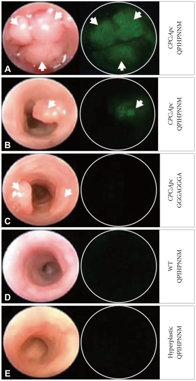
Fig. 1 Images under wide-field endoscopy videos after topical administration of fluorescence-labeled peptides. The left and right columns represent frames from white light and fluorescence, respectively. (A) The fluorescent-labeled target peptide FITC-Ahx-QPIHPNNM shows positive binding to multiple adenomas and (B) a single adenoma. (C) The control peptide shows minimal binding. (D) The target peptide also shows minimal binding to the lumen of the control mouse lacking Cre recombinase transgene and (E) the hyperplastic epithelium in a mutant K-ras mouse model. White arrows identify adenomas. Adapted from Miller et al. PLoS One 2011;6:e17384.21
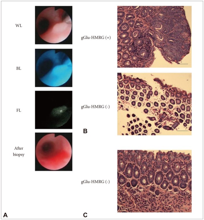
Fig. 2 γ-Glutamyl hydroxymethyl rhodamine green (gGlu-HMRG) fluorescence-guided tissue biopsy confirmed the presence of neoplastic lesions histologically. (A) gGlu-HMRG fluorescence-guided tissue biopsy under fluorescence colonoscopy. (B) Histology obtained by tissue biopsy. (C) Chronic microscopic colitis did not show detectable gGlu-HMRG fluorescence. Adapted from Mitsunaga et al. Gut 2013;62:1179-1186, with permission from BMJ Publishing Group Ltd.26 WL, white light image; BL, blue excitation light image; FL, fluorescence image.
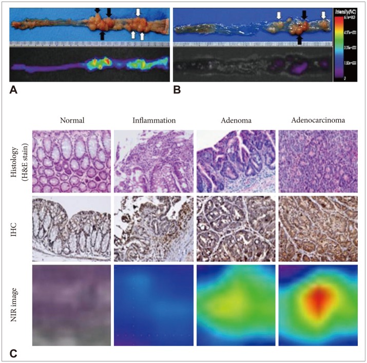
Fig. 3 Histology and near-infrared fluorescence (NIRF) imaging using an matrix metalloproteinase (MMP)-activatable probe in the AOM/DSS model. (A, B) Representative images of colons from BALB/c mice treated with AOM/DSS after injecting an MMP-activatable probe (A) or normal saline (B). (C) Histology and NIRF findings in BALB/c mice treated with AOM/DSS. Adapted from Yoon et al. Gut Liver 2010;4:488-497.29 IHC, immunohistochemistry; NIR, near infrared.
Reference
-
1. Yalamarthi S, Witherspoon P, McCole D, Auld CD. Missed diagnoses in patients with upper gastrointestinal cancers. Endoscopy. 2004; 36:874–879. PMID: 15452783.
Article2. van Rijn JC, Reitsma JB, Stoker J, Bossuyt PM, van Deventer SJ, Dekker E. Polyp miss rate determined by tandem colonoscopy: a systematic review. Am J Gastroenterol. 2006; 101:343–350. PMID: 16454841.
Article3. Matloff JL, Abidi W, Richards-Kortum R, Sauk J, Anandasabapathy S. High-resolution and optical molecular imaging for the early detection of colonic neoplasia. Gastrointest Endosc. 2011; 73:1263–1273. PMID: 21628019.
Article4. Dacosta RS, Wilson BC, Marcon NE. New optical technologies for earlier endoscopic diagnosis of premalignant gastrointestinal lesions. J Gastroenterol Hepatol. 2002; 17(Suppl):S85–S104. PMID: 12000596.
Article5. Goetz M, Wang TD. Molecular imaging in gastrointestinal endoscopy. Gastroenterology. 2010; 138:828–833. PMID: 20096697.
Article6. Uedo N, Iishi H, Tatsuta M, et al. A novel videoendoscopy system by using autofluorescence and reflectance imaging for diagnosis of esophagogastric cancers. Gastrointest Endosc. 2005; 62:521–528. PMID: 16185965.
Article7. Curvers WL, van Vilsteren FG, Baak LC, et al. Endoscopic trimodal imaging versus standard video endoscopy for detection of early Barrett's neoplasia: a multicenter, randomized, crossover study in general practice. Gastrointest Endosc. 2011; 73:195–203. PMID: 21168835.
Article8. Shahid MW, Buchner AM, Coron E, et al. Diagnostic accuracy of probe-based confocal laser endomicroscopy in detecting residual colorectal neoplasia after EMR: a prospective study. Gastrointest Endosc. 2012; 75:525–533. PMID: 22051243.
Article9. Shahid MW, Buchner AM, Heckman MG, et al. Diagnostic accuracy of probe-based confocal laser endomicroscopy and narrow band imaging for small colorectal polyps: a feasibility study. Am J Gastroenterol. 2012; 107:231–239. PMID: 22068663.
Article10. Shahid MW, Buchner AM, Raimondo M, Woodward TA, Krishna M, Wallace MB. Accuracy of real-time vs. blinded offline diagnosis of neoplastic colorectal polyps using probe-based confocal laser endomicroscopy: a pilot study. Endoscopy. 2012; 44:343–348. PMID: 22382851.
Article11. Goetz M, Malek NP, Kiesslich R. Microscopic imaging in endoscopy: endomicroscopy and endocytoscopy. Nat Rev Gastroenterol Hepatol. 2013; 7. 30. Epub. DOI: 10.1038/nrgastro.2013.134.
Article12. Goetz M, Watson A, Kiesslich R. Confocal laser endomicroscopy in gastrointestinal diseases. J Biophotonics. 2011; 4:498–508. PMID: 21567975.
Article13. Wallace MB, Fockens P. Probe-based confocal laser endomicroscopy. Gastroenterology. 2009; 136:1509–1513. PMID: 19328799.
Article14. Kiesslich R, Burg J, Vieth M, et al. Confocal laser endoscopy for diagnosing intraepithelial neoplasias and colorectal cancer in vivo. Gastroenterology. 2004; 127:706–713. PMID: 15362025.
Article15. Meining A, Saur D, Bajbouj M, et al. In vivo histopathology for detection of gastrointestinal neoplasia with a portable, confocal miniprobe: an examiner blinded analysis. Clin Gastroenterol Hepatol. 2007; 5:1261–1267. PMID: 17689297.
Article16. Wallace MB, Meining A, Canto MI, et al. The safety of intravenous fluorescein for confocal laser endomicroscopy in the gastrointestinal tract. Aliment Pharmacol Ther. 2010; 31:548–552. PMID: 20002025.
Article17. Schachschal G, Mayr M, Treszl A, et al. Endoscopic versus histological characterisation of polyps during screening colonoscopy. Gut. 2013; 6. 28. Epub. DOI: 10.1136/gutjnl-2013-304562.
Article18. Atreya R, Goetz M. Molecular imaging in gastroenterology. Nat Rev Gastroenterol Hepatol. Epub 2013 Jul 16. DOI: 10.1038/nrgastro.2013.125.
Article19. Marten K, Bremer C, Khazaie K, et al. Detection of dysplastic intestinal adenomas using enzyme-sensing molecular beacons in mice. Gastroenterology. 2002; 122:406–414. PMID: 11832455.
Article20. Li M, Wang TD. Targeted endoscopic imaging. Gastrointest Endosc Clin N Am. 2009; 19:283–298. PMID: 19423025.
Article21. Miller SJ, Joshi BP, Feng Y, Gaustad A, Fearon ER, Wang TD. In vivo fluorescence-based endoscopic detection of colon dysplasia in the mouse using a novel peptide probe. PLoS One. 2011; 6:e17384. PMID: 21408169.
Article22. Joshi BP, Liu Z, Elahi SF, Appelman HD, Wang TD. Near-infrared-labeled peptide multimer functions as phage mimic for high affinity, specific targeting of colonic adenomas in vivo (with videos). Gastrointest Endosc. 2012; 76:1197–1206. PMID: 23022051.
Article23. Joshi BP, Miller SJ, Lee CM, Seibel EJ, Wang TD. Multispectral endoscopic imaging of colorectal dysplasia in vivo. Gastroenterology. 2012; 143:1435–1437. PMID: 23041325.
Article24. Miller SJ, Lee CM, Joshi BP, Gaustad A, Seibel EJ, Wang TD. Targeted detection of murine colonic dysplasia in vivo with flexible multispectral scanning fiber endoscopy. J Biomed Opt. 2012; 17:021103. PMID: 22463021.
Article25. Urano Y. Novel live imaging techniques of cellular functions and in vivo tumors based on precise design of small molecule-based 'activatable' fluorescence probes. Curr Opin Chem Biol. 2012; 16:602–608. PMID: 23149093.
Article26. Mitsunaga M, Kosaka N, Choyke PL, et al. Fluorescence endoscopic detection of murine colitis-associated colon cancer by topically applied enzymatically rapid-activatable probe. Gut. 2013; 62:1179–1186. PMID: 22698650.
Article27. Tung CH, Mahmood U, Bredow S, Weissleder R. In vivo imaging of proteolytic enzyme activity using a novel molecular reporter. Cancer Res. 2000; 60:4953–4958. PMID: 10987312.28. Bremer C, Bredow S, Mahmood U, Weissleder R, Tung CH. Optical imaging of matrix metalloproteinase-2 activity in tumors: feasibility study in a mouse model. Radiology. 2001; 221:523–529. PMID: 11687699.
Article29. Yoon SM, Myung SJ, Ye BD, et al. Near-infrared fluorescence imaging using a protease-specific probe for the detection of colon tumors. Gut Liver. 2010; 4:488–497. PMID: 21253297.
Article30. Yoon SM, Myung SJ, Kim IW, et al. Application of near-infrared fluorescence imaging using a polymeric nanoparticle-based probe for the diagnosis and therapeutic monitoring of colon cancer. Dig Dis Sci. 2011; 56:3005–3013. PMID: 21465144.
Article31. Kim BJ, Yang SK, Kim JS, et al. Trends of ulcerative colitis-associated colorectal cancer in Korea: a KASID study. J Gastroenterol Hepatol. 2009; 24:667–671. PMID: 19378391.
Article32. Murthy SK, Kiesslich R. Evolving endoscopic strategies for detection and treatment of neoplastic lesions in inflammatory bowel disease. Gastrointest Endosc. 2013; 77:351–359. PMID: 23317581.
Article33. Gounaris E, Martin J, Ishihara Y, et al. Fluorescence endoscopy of cathepsin activity discriminates dysplasia from colitis. Inflamm Bowel Dis. 2013; 19:1339–1345. PMID: 23591598.
Article34. Awais D, Siegel CA, Higgins PD. Modelling dysplasia detection in ulcerative colitis: clinical implications of surveillance intensity. Gut. 2009; 58:1498–1503. PMID: 19651634.
Article35. Neumann H, Kiesslich R. Endomicroscopy and endocytoscopy in IBD. Gastrointest Endosc Clin N Am. 2013; 23:695–705. PMID: 23735111.
Article36. Neumann H, Vieth M, Günther C, et al. Virtual chromoendoscopy for prediction of severity and disease extent in patients with inflammatory bowel disease: a randomized controlled study. Inflamm Bowel Dis. 2013; 19:1935–1942. PMID: 23839228.37. Kiesslich R, Duckworth CA, Moussata D, et al. Local barrier dysfunction identified by confocal laser endomicroscopy predicts relapse in inflammatory bowel disease. Gut. 2012; 61:1146–1153. PMID: 22115910.
Article38. Van Cutsem E, Köhne CH, Láng I, et al. Cetuximab plus irinotecan, fluorouracil, and leucovorin as first-line treatment for metastatic colorectal cancer: updated analysis of overall survival according to tumor KRAS and BRAF mutation status. J Clin Oncol. 2011; 29:2011–2019. PMID: 21502544.
Article39. Goetz M, Hoetker MS, Diken M, Galle PR, Kiesslich R. In vivo molecular imaging with cetuximab, an anti-EGFR antibody, for prediction of response in xenograft models of human colorectal cancer. Endoscopy. 2013; 45:469–477. PMID: 23580409.
Article40. Thapa N, Kim S, So IS, et al. Discovery of a phosphatidylserine-recognizing peptide and its utility in molecular imaging of tumour apoptosis. J Cell Mol Med. 2008; 12(5A):1649–1660. PMID: 18363834.
Article
