Clin Endosc.
2014 Jan;47(1):74-78.
Observer Variability in Gastric Neoplasm Assessment Using the Vessel Plus Surface Classification for Magnifying Endoscopy with Narrow Band Imaging
- Affiliations
-
- 1Department of Internal Medicine, Kosin University College of Medicine, Busan, Korea. mipark@kosinmed.or.kr
Abstract
- BACKGROUND/AIMS
Recent studies have demonstrated that magnifying endoscopy with narrow band imaging (ME-NBI) facilitates differentiation of early gastric cancer from gastric adenoma using vessel plus surface (VS) classification. This study estimated the interobserver and intraobserver agreement of endoscopists using the Yao VS classification system for the gastric mucosal surface.
METHODS
We retrospectively reviewed patients who underwent endoscopic submucosal dissection or endoscopic mucosal resection, and selected cases in which preoperative ME-NBI was conducted. Before testing endoscopists, a 20-minute training module was given. Static ME-NBI images (n=47 cases) were presented to seven endoscopists (two experts and five trainees) who were asked to assess the images in 20 seconds using the Yao VS classification system. After 2 weeks, the endoscopists were asked to analyze the images again. The kappa statistic was calculated for intraobserver and interobserver variability.
RESULTS
The mean kappa for intraobserver agreement was 0.69 (experts, 0.74; trainees, 0.64). The mean kappa for interobserver agreement was 0.42 (experts, 0.49; trainees, 0.40).
CONCLUSIONS
We obtained reliable results as assessed by observer variability, with only brief training on VS classification. The VS classification appears to provide an objective assessment of ME-NBI for trainees who are not familiar with ME-NBI.
MeSH Terms
Figure
-
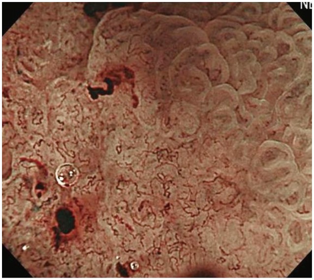
Fig. 1 Magnifying endoscopy with narrow band imaging showing a typical finding of demarcation line and irregular microvascular pattern.
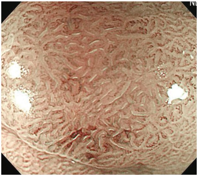
Fig. 2 Magnifying endoscopy with narrow band imaging showing a typical finding of regular microvascular and microsurface patterns
Reference
-
1. Miwa K, Doyama H, Ito R, et al. Can magnifying endoscopy with narrow band imaging be useful for low grade adenomas in preoperative biopsy specimens? Gastric Cancer. 2012; 15:170–178. PMID: 22407064.
Article2. Yao K. How is the VS (vessel plus surface) classification system applicable to magnifying narrow-band imaging examinations of gastric neoplasias initially diagnosed as low-grade adenomas? Gastric Cancer. 2012; 15:118–120. PMID: 22407063.
Article3. Gono K, Obi T, Yamaguchi M, et al. Appearance of enhanced tissue features in narrow-band endoscopic imaging. J Biomed Opt. 2004; 9:568–577. PMID: 15189095.
Article4. Nakayoshi T, Tajiri H, Matsuda K, Kaise M, Ikegami M, Sasaki H. Magnifying endoscopy combined with narrow band imaging system for early gastric cancer: correlation of vascular pattern with histopathology (including video). Endoscopy. 2004; 36:1080–1084. PMID: 15578298.
Article5. Yao K, Iwashita A, Tanabe H, et al. White opaque substance within superficial elevated gastric neoplasia as visualized by magnification endoscopy with narrow-band imaging: a new optical sign for differentiating between adenoma and carcinoma. Gastrointest Endosc. 2008; 68:574–580. PMID: 18656862.
Article6. Tsuji Y, Ohata K, Sekiguchi M, et al. Magnifying endoscopy with narrow-band imaging helps determine the management of gastric adenomas. Gastric Cancer. 2012; 15:414–418. PMID: 22252155.
Article7. Yao K, Anagnostopoulos GK, Ragunath K. Magnifying endoscopy for diagnosing and delineating early gastric cancer. Endoscopy. 2009; 41:462–467. PMID: 19418401.
Article8. Schlemper RJ, Riddell RH, Kato Y, et al. The Vienna classification of gastrointestinal epithelial neoplasia. Gut. 2000; 47:251–255. PMID: 10896917.
Article9. Viera AJ, Garrett JM. Understanding interobserver agreement: the kappa statistic. Fam Med. 2005; 37:360–363. PMID: 15883903.10. Kim YJ, Park JC, Kim JH, et al. Histologic diagnosis based on forceps biopsy is not adequate for determining endoscopic treatment of gastric adenomatous lesions. Endoscopy. 2010; 42:620–626. PMID: 20623445.
Article11. Yao K, Iwashita A, Tanabe H, et al. Novel zoom endoscopy technique for diagnosis of small flat gastric cancer: a prospective, blind study. Clin Gastroenterol Hepatol. 2007; 5:869–878. PMID: 17544872.
Article12. Kaise M, Kato M, Urashima M, et al. Magnifying endoscopy combined with narrow-band imaging for differential diagnosis of superficial depressed gastric lesions. Endoscopy. 2009; 41:310–315. PMID: 19340733.
Article13. Miwa H, Yokoyama T, Hori K, et al. Interobserver agreement in endoscopic evaluation of reflux esophagitis using a modified Los Angeles classification incorporating grades N and M: a validation study in a cohort of Japanese endoscopists. Dis Esophagus. 2008; 21:355–363. PMID: 18477259.
Article14. Nasseri-Moghaddam S, Razjouyan H, Nouraei M, et al. Inter- and intra-observer variability of the Los Angeles classification: a reassessment. Arch Iran Med. 2007; 10:48–53. PMID: 17198454.
- Full Text Links
-
- Actions
-
Cited
- CITED
-
- Close
- Share
-
- Similar articles
-
- Usefulness of Narrow-Band Imaging in Endoscopic Submucosal Dissection of the Stomach
- Image-Enhanced Endoscopy and Its Corresponding Histopathology in the Stomach
- Clinical Application of Magnifying Endoscopy with Narrow-Band Imaging in the Stomach
- Clinical Role of Magnifying Endoscopy with Narrow-band Imaging in the Diagnosis of Early Gastric Cancer
- Application of artificial intelligence for diagnosis of early gastric cancer based on magnifying endoscopy with narrow-band imaging
