Endoscopic Ultrasound-Guided Sampling of a Metastatic Mucinous Adenocarcinoma Mimicking a Gastric Subepithelial Tumor
- Affiliations
-
- 1Department of Internal Medicine, Digestive Disease Center, Soonchunhyang University Hospital, Soonchunhyang University College of Medicine, Seoul, Korea.
- 2Institute for Digestive Research, Digestive Disease Center, Soonchunhyang University Hospital, Soonchunhyang University College of Medicine, Seoul, Korea. iman0825@naver.com
- 3Department of Pathology, Soonchunhyang University College of Medicine, Seoul, Korea.
- 4Department of Oncology, Soonchunhyang University Hospital, Soonchunhyang University College of Medicine, Seoul, Korea.
- KMID: 2165381
- DOI: http://doi.org/10.5946/ce.2014.47.5.460
Abstract
- Metastatic mucinous adenocarcinoma of appendix origin and mimicking a gastric subepithelial tumor (SET) is very rare. Endoscopic ultrasound (EUS)-guided sampling is a useful diagnostic method for SETs. However, the cytologic findings of metastatic mucinous adenocarcinoma are unfamiliar to many pathologists and gastroenterologists. These findings present a diagnostic challenge because the introduction of gastric epithelium and mucin into the specimen during the procedure can be misleading. This is the first reported experience of an EUS-guided sampling of a gastric SET in a patient with suspected appendiceal tumor, to make the diagnosis of a mucinous adenocarcinoma.
MeSH Terms
Figure
-
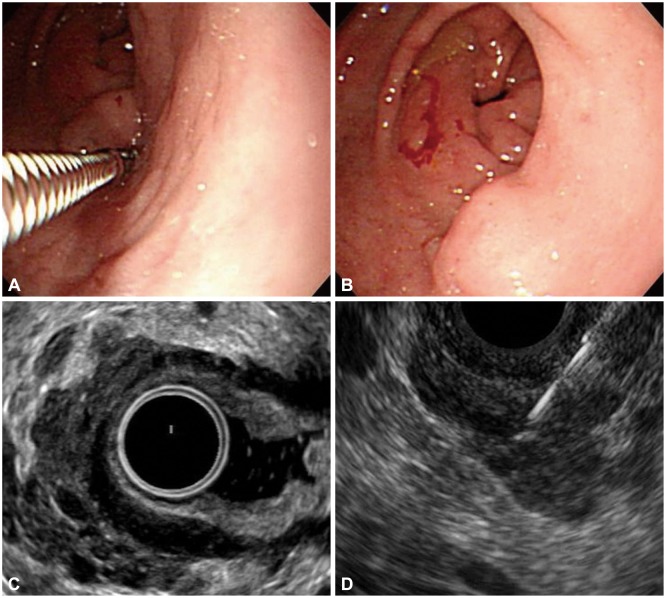
Fig. 1 Endoscopy, endoscopic ultrasound (EUS), and EUS-guided sampling of the gastric subepithelial tumor (SET). (A) Posterior portion of a dumbbell-shaped SET is noted on the antrum with a normal overlying mucosa. (B) Forceps biopsy is performed in the linear ulcer on the surface of anterior portion of the SET. (C) The hypoechoic mass with an amorphous shape has invaded into the gastric wall layers, and is accompanied with multiple anechoic portions with variable sizes. (D) EUS-guided ProCore needle biopsy is performed on the lesion.
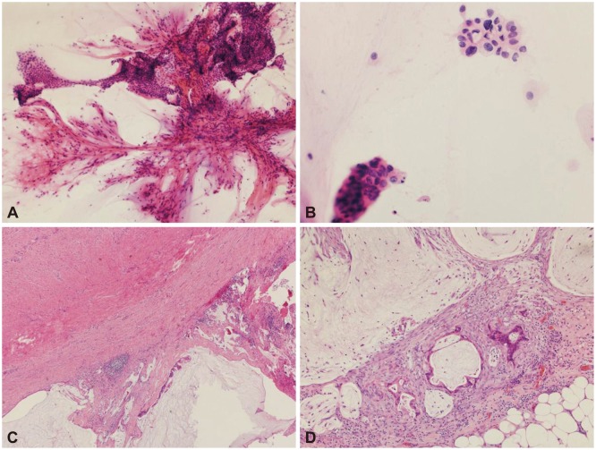
Fig. 2 Pathologic findings of the gastric subepithelial tumor. (A) Lower-power view of the endoscopic ultrasound-guided sampling depicts several branching vasculatures with folded sheets of bland-looking mucinous epithelial cells (H&E stain, ×100). (B) Some of the mucinous cells show nuclear pleomorphism and hyperchromasia, suspicious for malignancy (H&E stain, ×400). (C) The serosa of the resected appendix shows extension of the mucinous adenocarcinoma with several acellular mucin pools (H&E stain, ×40). (D) There are a few glands of metastatic mucinous adenocarcinoma with surrounding inflammation and fibrosis in the omentum. Several acellular mucin polls are also seen (H&E stain, ×100).
Reference
-
1. Holland JF, Frei E, Bast RC, Morton DL. Tumours of appendix. In : Holland JF, Frei E, Bast RC, Morton DL, editors. Cancer Medicine. 3rd ed. Philadelphia: Lea & Febiger;1993.2. Lee NK, Kim S, Kim GH, et al. Hypervascular subepithelial gastrointestinal masses: CT-pathologic correlation. Radiographics. 2010; 30:1915–1934. PMID: 21057127.
Article3. Que Y, Tao C, Wang X, Zhang Y, Chen B. Pseudomyxoma peritonei: some different sonographic findings. Abdom Imaging. 2012; 37:843–848. PMID: 22234650.
Article4. Peter S, Eltoum I, Eloubeidi MA. EUS-guided FNA of peritoneal carcinomatosis in patients with unknown primary malignancy. Gastrointest Endosc. 2009; 70:1266–1270. PMID: 19640520.
Article5. Rana SS, Bhasin DK, Gupta R, Singh K. EUS-guided FNA of peritoneal carcinomatosis. Gastrointest Endosc. 2011; 73:188–189. PMID: 21184887.
Article6. Dumonceau JM, Polkowski M, Larghi A, et al. Indications, results, and clinical impact of endoscopic ultrasound (EUS)-guided sampling in gastroenterology: European Society of Gastrointestinal Endoscopy (ESGE) Clinical Guideline. Endoscopy. 2011; 43:897–912. PMID: 21842456.
Article
- Full Text Links
-
- Actions
-
Cited
- CITED
-
- Close
- Share
-
- Similar articles
-
- Subepithelial Tumor-like Gastric Cancer
- Tuberculous Lymphadenitis Mimicking Gastric Subepithelial Tumor Diagnosed Using Endoscopic Ultrasound-guided Fine-needle Aspiration
- Metastatic Mucinous Adenocarcinoma Mimicking Cerebral Hemorrhage
- Two cases of mucinous adenocarcinoma of the stomach mistaken as submucosal tumor
- Endoscopic Management of Gastric Subepithelial Tumor
