Clin Endosc.
2014 Sep;47(5):452-454. 10.5946/ce.2014.47.5.452.
Esophageal Involvement of Pemphigus Vulgaris Associated with Upper Gastrointestinal Bleeding
- Affiliations
-
- 1Department of Internal Medicine, Yonsei University College of Medicine, Seoul, Korea. sjpark@yuhs.ac
- 2Institute of Gastroenterology, Yonsei University College of Medicine, Seoul, Korea.
- KMID: 2165379
- DOI: http://doi.org/10.5946/ce.2014.47.5.452
Abstract
- Esophageal involvement of pemphigus vulgaris is rare, and when present, the most common presenting symptoms reported in the medical literature are odynophagia and dysphagia. Here, we present two cases of pemphigus vulgaris presenting with upper gastrointestinal hemorrhage because of esophageal involvement of the disease. In case 1, a 41-year-old female patient with a prior diagnosis of pemphigus vulgaris presented with hematemesis. Esophagogastroduodenoscopy showed diffuse mucosal exfoliation and oozing bleeding of the oropharynx and esophagus. The patient recovered after the administration of high-dose corticosteroids and immunosuppressants. In case 2, a 30-year-old female patient with known pemphigus vulgaris also presented with hematemesis, showing similar endoscopic findings to the first case. She also responded to the same treatment. Esophageal involvement of pemphigus vulgaris responds to high-dose corticosteroids and immunosuppressants. Thus, in patients with pemphigus vulgaris with signs or symptoms of upper gastrointestinal bleeding, an early endoscopy for the evaluation of esophageal involvement is beneficial.
MeSH Terms
Figure
-
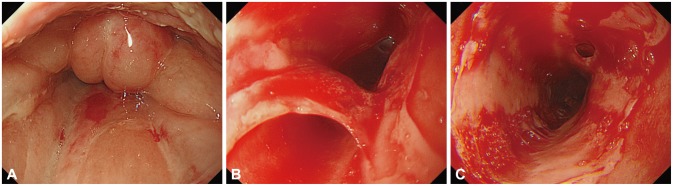
Fig. 1 Endoscopic findings. (A) Diffuse swelling of the larynx with oral desquamation. (B) Multiple erosions and shallow ulcers with exfoliation of the esophageal mucosal tissue. (C) Diffuse subepithelial hemorrhage of the esophagus.
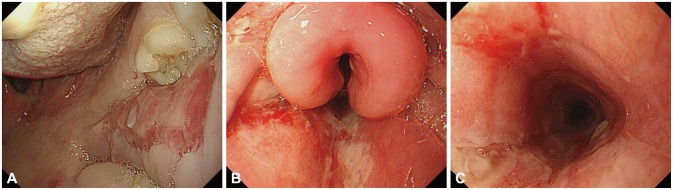
Fig. 2 Endoscopic findings. (A) Mucosal abrasions on the oral cavity wall. (B) Erythematous mucosa with severe edema of the larynx. (C) Linear ulceration in the esophagus covered with exudate.
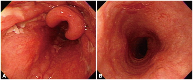
Fig. 3 Endoscopic findings. (A) Improvement of laryngeal edema and oral mucosal lesions. (B) Resolved status of mucosal lesions in the esophagus.
Reference
-
1. Mignogna MD, Lo Muzio L, Galloro G, Satriano RA, Ruocco V, Bucci E. Oral pemphigus: clinical significance of esophageal involvement: report of eight cases. Oral Surg Oral Med Oral Pathol Oral Radiol Endod. 1997; 84:179–184. PMID: 9269021.2. Kaplan RP, Touloukian J, Ahmed AR, Newcomer VD. Esophagitis dissecans superficialis associated with pemphigus vulgaris. J Am Acad Dermatol. 1981; 4:682–687. PMID: 7016940.
Article3. Schissel DJ, David-Bajar K. Esophagitis dissecans superficialis associated with pemphigus vulgaris. Cutis. 1999; 63:157–160. PMID: 10190066.4. Galloro G, Mignogna M, de Werra C, et al. The role of upper endoscopy in identifying oesophageal involvement in patients with oral pemphigus vulgaris. Dig Liver Dis. 2005; 37:195–199. PMID: 15888285.
Article5. Faias S, Lage P, Sachse F, et al. Pemphigus vulgaris with exclusive involvement of the esophagus: case report and review. Gastrointest Endosc. 2004; 60:312–315. PMID: 15278072.
Article
- Full Text Links
-
- Actions
-
Cited
- CITED
-
- Close
- Share
-
- Similar articles
-
- Combined Upper Gastrointestinal Lesions with Esophageal Varices
- A Case of Esophageal Involvement in Pemphigus Vulgaris
- Persistent Pemphigus Vulgaris Showing Features of Tufted Hair Folliculitis
- A Case Report of Paraneoplastic Pemphigus Associated with Esophageal Squamous Cell Carcinoma
- A Case of Pemphigus Vulgaris
