Various Upper Endoscopic Findings of Acute Esophageal Thermal Injury Induced by Diverse Food: A Case Series
- Affiliations
-
- 1Department of Internal Medicine, Konyang University College of Medicine, Daejeon, Korea. ismkim@kyuh.ac.kr
- KMID: 2165378
- DOI: http://doi.org/10.5946/ce.2014.47.5.447
Abstract
- Esophageal thermal injury caused by food has been reported to occur mostly after drinking hot liquid food, and is known to produce alternating white and red linear mucosal bands. In addition, thermal injury caused by ingestion of hot solid foods is documented to be a cause of esophageal ulcers or pseudomembranes. From January 2006 to August 2012, five patients with suspected esophageal thermal injury underwent esophagogastroduodenoscopy with biopsy. A "candy-cane" appearance was observed in one case, pseudomembrane was observed in two cases, an esophageal ulcer was observed in one case, and a friable and edematous mucosa was noted in one case. We believe that the endoscopic findings of esophageal thermal injury depend on the following factors: causative materials, amount of food consumed, exposure period, and time to endoscopy after the incident. Therefore, physicians who encounter patients with suspected esophageal thermal injury should carefully take the patient's history considering these factors.
Figure
-
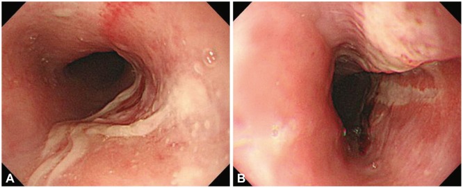
Fig. 1 Endoscopic findings. (A) The initial endoscopic view shows whitish pseudomembranous mucosa with a geographic shape. (B) Follow-up endoscopic findings after 7 days. The candy-cane appearance is seen.
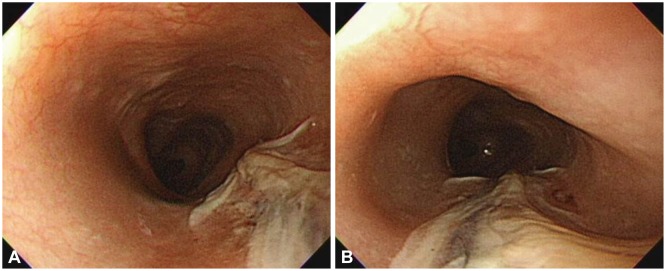
Fig. 2 (A, B) Endoscopic findings. The initial endoscopic view shows a whitish pseudomembrane extending from the proximal third of the esophagus to the distal portion.
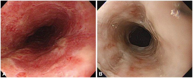
Fig. 3 Endoscopic findings. (A) The initial endoscopic view shows a whitish pseudomembrane that nearly completely encircles the esophageal mucosa. (B) Follow-up endoscopic findings after 7 days. Hyperemic and edematous mucosa and mild whitish fibrosis can be seen on the upper and middle esophagus.

Fig. 4 (A, B) Endoscopic findings, showing an edematous, friable esophageal mucosa that bleeds easily on touching.
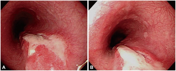
Fig. 5 (A, B) Endoscopic findings. A large 3.5×2.5 cm ulcer with a severely erythematous friable mucosa is seen noted 34 cm below the incisor.
Reference
-
2. Chung WC, Paik CN, Jung JH, Kim JD, Lee KM, Yang JM. Acute thermal injury of the esophagus from solid food: clinical course and endoscopic findings. J Korean Med Sci. 2010; 25:489–491. PMID: 20191054.
Article3. Dutta SK, Chung KY, Bhagavan BS. Thermal injury of the esophagus. N Engl J Med. 1998; 339:480–481. PMID: 9705693.
Article4. Lieberman DA, Keeffe EB. Esophageal burn and the microwave oven. Ann Intern Med. 1982; 97:137. PMID: 7091986.
Article5. Eliakim R. Thermal injury from a hamburger: a rare cause of odynophagia. Gastrointest Endosc. 1999; 50:282–283. PMID: 10425431.
Article6. Choi EK, Lee GH, Jung HY, et al. The healing course of thermal esophageal injury: a case report. Gastrointest Endosc. 2005; 62:158–160. PMID: 15990841.
Article7. Kim SM, Jung SH, Kim YM, et al. Esophageal ulcer by thermal injury from steamed egg. Korean J Gastrointest Endosc. 2006; 32:190–193.
- Full Text Links
-
- Actions
-
Cited
- CITED
-
- Close
- Share
-
- Similar articles
-
- A Large Symmetrical Esophageal Ulcer Caused by Thermal and Compressive Injury from a Solid Foodstuff Known as 'Song-Pyen'
- A Case of PICOLIGHT Powder Induced Thermal Injury of the Gastric Mucosa
- Acute Thermal Injury of the Esophagus from Solid Food: Clinical Course and Endoscopic Findings
- Esophageal Ulcer by Thermal Injury from Steamed Egg
- Physiology of Upper Esophageal Sphincter Contraction
