Large Mature Cystic Teratoma of the Third Ventricle in Infancy: A Case Report and Review of Literatures
- Affiliations
-
- 1Department of Neurosurgery, Hanyang University Medical Center, Seoul, Korea. ksy8498@hanyang.ac.kr
- 2Department of Neurosurgery, Hanyang University Guri Hospital, Guri, Korea.
- KMID: 2165235
- DOI: http://doi.org/10.14791/btrt.2016.4.1.44
Abstract
- Teratomas of the central nervous system are rare and are frequently found in children and young adults. Cystic teratomas found in infancy is a well-recognized but infrequent entity. Intracranial teratomas,like teratomas in general, tend to arise from midline structures such as the pineal gland, but has rarely been found in the third ventricle. We report a rare case of a 6-month-old infant with a mature cystic teratoma of the third ventricle with a review of literatures
Keyword
Figure
-
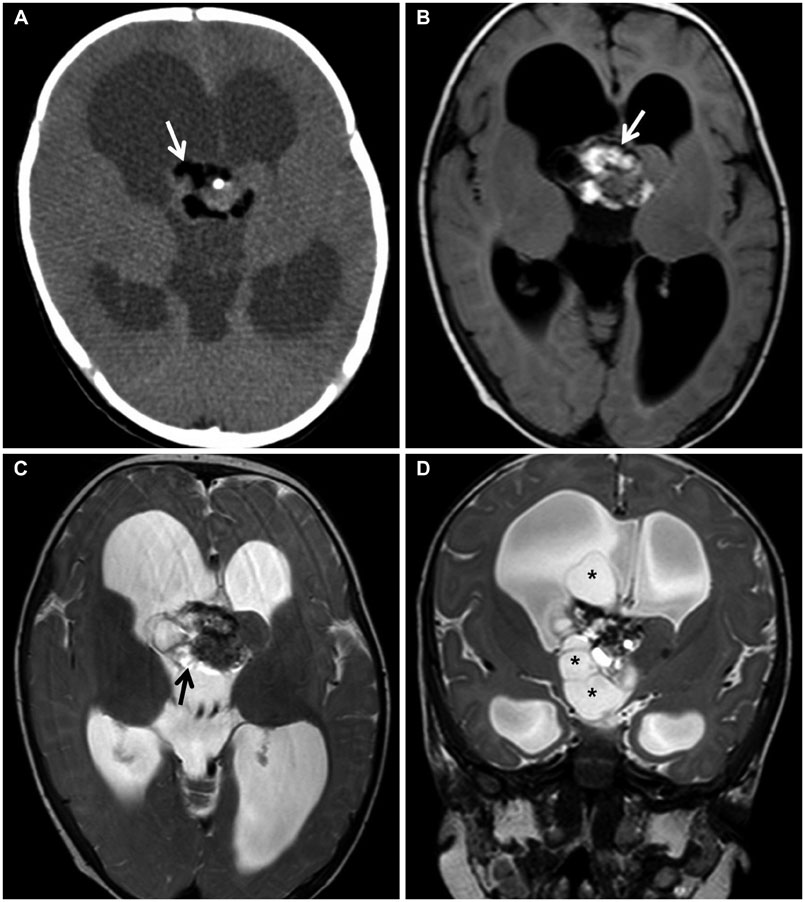
Fig. 1 A: Preoperative CT scan reveals a mass in the third ventricle with extension to the right lateral ventricle, consisting of fat components (white arrow). B and C: Preoperative magnetic resonance (MR) axial image shows a heterogenous mass with portions of calcification (white arrow) and fat tissue (black arrow), suggesting a teratoma. D: Preoperative MR coronal image shows a multi-cystic mass (asterisks) involving the third and right lateral ventricles.

Fig. 2 A: Gross view of the tumor specimen shows a grayish white soft tissue, measuring 2.8×2.5×2 cm in dimensions. B: Gross view of the cut surface of the tumor specimen shows homogenous tan color and focal calcification.

Fig. 3 A: Low power photographic veiw of the tumor showing a mixture of bronchial lining epithelium, bronchial glands, muscle, adipose tissue and squamous cell lining cyst filled with keratin materials (hematoxylin-eosin, ×20). B: Representitive view of the tumor components consist of bronchial repiratory epithelium and bronchial cartilage (hematoxylin-eosin, ×100)
Reference
-
1. Goyal N, Singh PK, Kakkar A, Sharma MC, Mahapatra AK. Mature teratoma in association with neural tube defect (occipital encephalocele): series of four cases and review of the literature. Pediatr Neurosurg. 2012; 48:67–72.
Article2. Sinha VD, Dharker SR, Pandey CL. Congenital intracranial teratoma of the lateral ventricle. Neurol India. 2001; 49:170–173.3. Zhou P, Li Y, Yang Z, Shu J. Mature cystic teratoma of skull base and attached to the wall of third ventricle. Turk Neurosurg. 2014; 24:292–293.4. Echevarria ME, Fangusaro J, Goldman S. Pediatric central nervous system germ cell tumors: a review. Oncologist. 2008; 13:690–699.
Article5. Jubran RF, Finlay J. Central nervous system germ cell tumors: controversies in diagnosis and treatment. Oncology (Williston Park). 2005; 19:705–711. discussion 711-2, 715-7, 721.6. Agrawal M, Uppin MS, Patibandla MR, et al. Teratomas in central nervous system: a clinico-morphological study with review of literature. Neurol India. 2010; 58:841–846.
Article7. Verma R, Malone S, Canil C, Jansen G, Lesiuk H. Primary skull-based yolk-sac tumour: case report and review of central nervous system germ cell tumours. J Neurooncol. 2011; 101:129–134.
Article8. Sandow BA, Dory CE, Aguiar MA, Abuhamad AZ. Best cases from the AFIP: congenital intracranial teratoma. Radiographics. 2004; 24:1165–1170.9. Kim JM, Cheong JH, Yi HJ, Bak KH, Kim CH, Oh SJ. Metachronous germinoma after total removal of mature teratoma in the third ventricle: a case report. J Korean Med Sci. 2002; 17:287–291.
Article10. Goyal N, Kakkar A, Singh PK, et al. Intracranial teratomas in children: a clinicopathological study. Childs Nerv Syst. 2013; 29:2035–2042.
Article11. Rosenblum MK, Nakazato Y, Matsutani M. CNS germ cell tumours. In : Louis DN, Ohgaki H, Wiestler OD, Cavenee WK, editors. WHO classification of tumours of the central nervous system. 4th ed. IARC: Lyon;2007. p. 197–204.12. Tobias S, Valarezo J, Meir K, Umansky F. Giant cavernous sinus teratoma: a clinical example of a rare entity: case report. Neurosurgery. 2001; 48:1367–1370. discussion 1370-1.
Article13. Sano K. Pathogenesis of intracranial germ cell tumors reconsidered. J Neurosurg. 1999; 90:258–264.
Article
- Full Text Links
-
- Actions
-
Cited
- CITED
-
- Close
- Share
-
- Similar articles
-
- A Case of Adenocarcinoma Arising in Mature Cystic Teratoma of the Ovary
- A Case of Papillary Carcinoma of Thyroid Gland Arising from Ovarian Mature Cystic Teratoma
- A Case of Anaplastic Carcinoma Arising from Mature Cystic Teratoma of Ovary
- A Case of Bilateral Testicular Teratoma Found in Infant
- A Case of Delayed Operated Huge Mature Cystic Teratoma of the Ovary
