Brain Tumor Res Treat.
2013 Apr;1(1):54-56. 10.14791/btrt.2013.1.1.54.
Pituitary Symptomatic Salivary Gland Rest Cyst: Case Report
- Affiliations
-
- 1Department of Neurosurgery, Kyungpook National University, Daegu, Korea. jhwang@knu.ac.kr
- KMID: 2165225
- DOI: http://doi.org/10.14791/btrt.2013.1.1.54
Abstract
- Although salivary gland rest in posterior pituitary is frequently observed during microscopic examination at autopsy, this rest tissue rarely causes clinical symptoms. So far, only three symptomatic salivary gland rest cyst in pituitary was reported. The author reports the 4th case of symptomatic pituitary salivary gland rest cyst in 26-year-old woman presenting with severe headache. The lesion showed hyperdense shadow on computed tomography, high signal on T1-weighted image and low signal on T2-weighted image magnetic resonance imaging. There was no enhancement of the mass after gadolinium injection. Trans-sphenoidal approach revealed that the cyst wall consisted of normal appearing salivary gland and gelatinous cyst content. Although the pituitary salivary gland rest cyst is rarely symptomatic, it should be take into consideration in the differential diagnosis of pituitary lesions.
MeSH Terms
Figure
-

Fig. 1 A: Computed tomography revealed hyperdense shadow on sella turcica. A small nodular lesion in posterior pituitary shows low signal on T2-weighted image (B) and high signal on T1-weighted image (C). D: There was no enhancement of the mass after gadolinium injection.
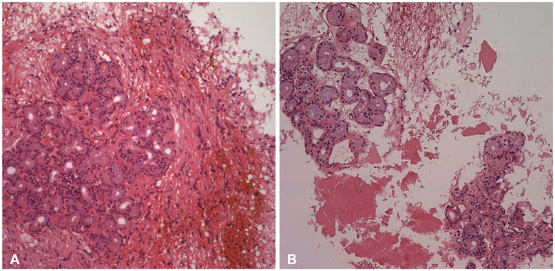
Fig. 2 A: Microscopic examination showed well-formed salivary acini with a low-columnar epithelium in a fibrovascular stroma in cyst wall. B: The eosinophilic secretions are consistent with the gelatinous material found at surgery (hematoxylin and eosin, ×40).
Reference
-
1. Schochet SS Jr, McCormick WF, Halmi NS. Salivary gland rests in the human pituitary. Light and electron microscopical study. Arch Pathol. 1974; 98:193–200.2. Chimelli L, Gadelha MR, Une K, et al. Intra-sellar salivary gland-like pleomorphic adenoma arising within the wall of a Rathke's cleft cyst. Pituitary. 2000; 3:257–261.3. Kato T, Aida T, Abe H, et al. [Ectopic salivary gland within the pituitary gland. Case report]. Neurol Med Chir (Tokyo). 1988; 28:930–933.4. Tatter SB, Edgar MA, Klibanski A, Swearingen B. Symptomatic salivary-rest cyst of the sella turcica. Acta Neurochir (Wien). 1995; 135:150–153.
Article5. Chen CH, Hsu SS, Lai PH, Lo YS. Intrasellar symptomatic salivary gland rest. J Chin Med Assoc. 2007; 70:215–217.
Article6. Dubois PM, el Amraoui A, Héritier AG. Development and differentiation of pituitary cells. Microsc Res Tech. 1997; 39:98–113.
Article7. Rodriguez F, Scheithauer BW, Ockner DM, Giannini C. Solitary fibrous tumor of the cerebellopontine angle with salivary gland heterotopia: a unique presentation. Am J Surg Pathol. 2004; 28:139–142.
Article8. Garrett JR, Kidd A. The innervation of salivary glands as revealed by morphological methods. Microsc Res Tech. 1993; 26:75–91.
Article9. Fager CA, Carter H. Intrasellar epithelial cysts. J Neurosurg. 1966; 24:77–81.
Article10. Asari S, Ito T, Tsuchida S, Tsutsui T. MR appearance and cyst content of Rathke cleft cysts. J Comput Assist Tomogr. 1990; 14:532–535.
Article
- Full Text Links
-
- Actions
-
Cited
- CITED
-
- Close
- Share
-
- Similar articles
-
- A Case Report of Symptomatic Salivary Gland Rest within the Pituitary Gland
- A case report of the salivary duct cyst and review of literatures
- Colloid Cyst in Pituitary Gland: A Case Report
- A Case of Ectopic Salivary Gland with Fistula in the Anterior Neck
- Multiple sialolithiasis in sublingual gland: report of a case
