An Intracranial Chondroma with Intratumoral Hemorrhage: A Case Report and Review of the Literature
- Affiliations
-
- 1Department of Neurosurgery, Seoul St. Mary's Hospital, The Catholic University of Korea College of Medicine, Seoul, Korea. ssjeun@catholic.ac.kr
- KMID: 2165222
- DOI: http://doi.org/10.14791/btrt.2013.1.1.42
Abstract
- A 55-year-old female presented to the emergency room with a complaint of aphasia. Her initial brain computed tomography scan showed an intracranial hemorrhage in the left frontal area. After surgery, histopathological examination confirmed the diagnosis of a chondroma. Intradural chondroma is a rare, slow growing, benign intracranial neoplasm, but is even rarer in combination with an intratumoral hemorrhage. Chondromas are generally avascular cartilaginous lesions. Our case was thought to be caused by the rupture of abnormally weak vessels derived from the friable tumor. Intradural chondromas may be included in the differential diagnosis of intracranial tumors with acute hemorrhages.
Keyword
MeSH Terms
Figure
-
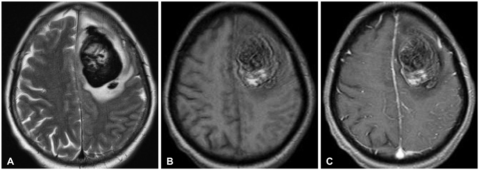
Fig. 1 Preoperative MRI images show a space-occupying lesion attached to the falx and dura at the left frontal area. A: T2-weighted image. B: T1-weighted image. C: T1-weighted enhanced image. MRI: magnetic resonance imaging.
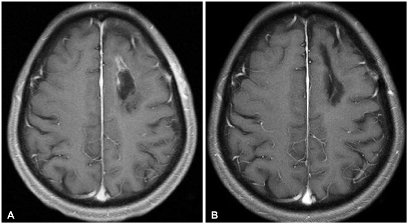
Fig. 2 Postoperative image study. A: The tumor was totally removed according to the postoperative MRI scan. B: A six month follow-up MRI scan showed no evidence of recurrence (both, T1-weighted enhanced image). MRI: magnetic resonance imaging.
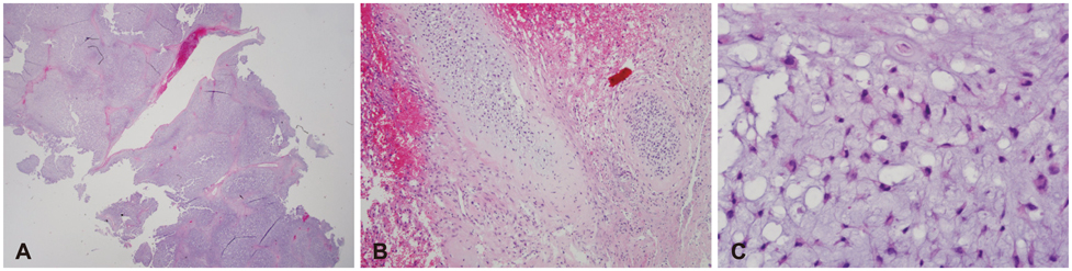
Fig. 3 Histologic finding of tumor specimen. A: An intracranial tumor, which showed a lobulated growth pattern (×40, H&E). B: Hemorrhagic areas were present inside the tumor (×100, H&E). C: Tumor cells were distributed in the myxoid cartilaginous matrix with eosinophilic cytoplasm and absence of nuclear atypism. A few tumor cells had lacunae (×400, H&E).
Reference
-
1. Linsen M, Junmei W, Liwei Z, Jianping D, Xuzhu C. An intracranial chondroma with intratumoral and subarachnoidal hemorrhage. Neurol India. 2011; 59:310–313.
Article2. Ghogawala Z, Moore M, Strand R, Kupsky WJ, Scott RM. Clival chondroma in a child with Ollier's disease Case report. Pediatr Neurosurg. 1991-1992; 17:53–56.3. Colpan E, Attar A, Erekul S, Arasil E. Convexity dural chondroma: a case report and review of the literature. J Clin Neurosci. 2003; 10:106–108.
Article4. Padhya TA, Athavale SM, Kathju S, Sarkar S, Mehta AR. Osteochondroma of the skull base. Otolaryngol Head Neck Surg. 2007; 137:166–168.
Article5. Bakdash H, Alksne JF, Rand RW. Osteochondroma of the base of the skull causing an isolated oculomotor nerve paralysis. Case report emphasizing microsurgical techniques. J Neurosurg. 1969; 31:230–233.
Article6. Khosrovi H, Sadrolhefazi A, el-Kadi H, Bloomfield SM, Schochet SS. Intradural convexity chondroma: a case report and review of diagnostic features. W V Med J. 2000; 96:612–616.7. Mapstone TB, Wongmongkolrit T, Roessman U, Ratcheson RA. Intradural chondroma: a case report and review of the literature. Neurosurgery. 1983; 12:111–114.
Article8. Matz S, Israeli Y, Shalit MN, Cohen ML. Computed tomography in intracranial supratentorial osteochondroma. J Comput Assist Tomogr. 1981; 5:109–115.
Article9. Esiri M. Russell and Rubinstein's pathology of tumors of the nervous system. Sixth edition. J Neurol Neurosurg Psychiatry. 2000; 68:538D.
Article10. Xin Y, Hao S, Zhang J, et al. Microsurgical treatment of intracranial chondroma. J Clin Neurosci. 2011; 18:1064–1071.
Article
- Full Text Links
-
- Actions
-
Cited
- CITED
-
- Close
- Share
-
- Similar articles
-
- Chondrosarcoma with Intratumoral Hemorrhage: Case Report
- Maffucci's Syndrome Complicated by an Intracranial Chondroma
- Intracranial Intradural Chondroma: Case Report
- Giant Extra-Capsular Synovial Chondroma of the knee joint: A Case Report
- A Case of Falx Meningioma Diagnosed due to Intratumoral Hemorrhage: Case Report
