Screening Ultrasound in Women with Negative Mammography: Outcome Analysis
- Affiliations
-
- 1Department of Radiology, Samsung Medical Center, Sungkyunkwan University School of Medicine, Seoul, Korea. bkhan@skku.edu
- 2Department of Radiology, Kangnam Sacred Heart Hospital, Hallym University College of Medicine, Seoul, Korea.
- KMID: 2163628
- DOI: http://doi.org/10.3349/ymj.2015.56.5.1352
Abstract
- PURPOSE
To show the results of an audit of screening breast ultrasound (US) in women with negative mammography in a single institution and to analyze US-detected cancers within a year and interval cancers.
MATERIALS AND METHODS
During the year of 2006, 1974 women with negative mammography were screened with US in our screening center, and 1727 among them had pathologic results or any follow up breast examinations more than a year. We analyzed the distribution of Breast Imaging Reporting and Data System (BI-RADS) category and the performance outcome through follow up.
RESULTS
Among 1727 women (age, 30-76 years, median 49.5 years), 1349 women (78.1%) showed dense breasts on mammography, 762 (44.1%) had previous breast US, and 25 women (1.4%) had a personal history of breast cancers. Test negatives were 94.2% (1.627/1727) [BI-RADS category 1 in 885 (51.2%), 2 in 742 (43.0%)]. The recall rate (=BI-RADS category 3, 4, 5) was 5.8%. Eight cancers were additionally detected with US (yield, 4.6 per 1000). The sensitivity, specificity, and positive predictive value (PPV1, PPV2) were 88.9%, 94.6%, 8.0%, and 28.0%, respectively. Eight of nine true positive cancers were stage I or in-situ cancers. One interval cancer was stage I cancer from BI-RADS category 2.
CONCLUSION
Screening US detected 4.6 additional cancers among 1000. The recall rate was 5.8%, which is in lower bound of acceptable range of mammography (5-12%), according to American College of Radiology standard.
MeSH Terms
Figure
-
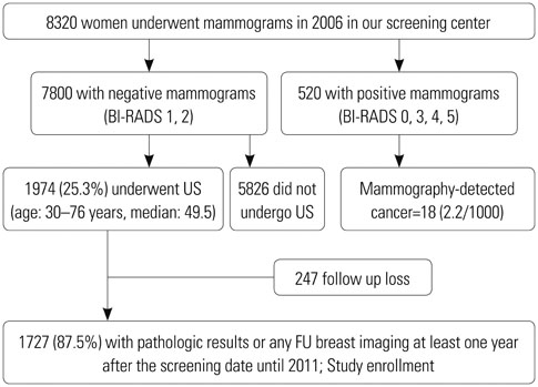
Fig. 1 Study population. BI-RADS, Breast Imaging Reporting and Data System; US, ultrasound; FU, follow up.
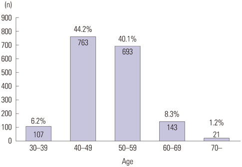
Fig. 2 Age distribution of study population.
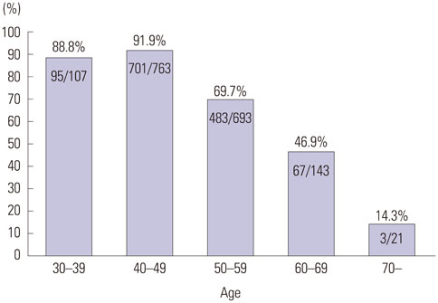
Fig. 3 Proportion of dense breasts according to the women's age.
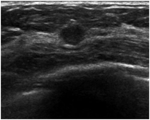
Fig. 4 A US-detected cancer in a 49-year-old woman that was initially assessed as BI-RADS category 4A (case 2 in Table 3). US shows an illdefined oval hypoechoic mass in right breast and it was assessed as category 4A. The pathologic diagnosis was invasive ductal cancer with ductal carcinoma in situ. US, ultrasound; BI-RADS, Breast Imaging Reporting and Data System.
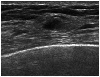
Fig. 5 A supplementary screening US detected cancer case in a 40-year-old woman that was initially assessed as BI-RADS category 3 (case 8 in Table 2). Initial supplementary screening US was assessed as BI-RADS 3 due to multiple small oval isoechoic nodules in both breasts. Diagnostic US after 6 months revealed that the mass in right breast increased in size up to 1.1 cm, and it was still not palpable. Both initial and follow-up mammograms were negative (not shown here). The pathologic diagnosis was invasive ductal cancer. US, ultrasound; BI-RADS, Breast Imaging Reporting and Data System.
Cited by 1 articles
-
Lymphangiogenesis in Breast Cancer Correlates with Matrix Stiffness on Shear-Wave Elastography
Yoon Jin Cha, Ji Hyun Youk, Baek Gil Kim, Woo Hee Jung, Nam Hoon Cho
Yonsei Med J. 2016;57(3):599-605. doi: 10.3349/ymj.2016.57.3.599.
Reference
-
1. Tabár L, Vitak B, Chen HH, Yen MF, Duffy SW, Smith RA. Beyond randomized controlled trials: organized mammographic screening substantially reduces breast carcinoma mortality. Cancer. 2001; 91:1724–1731.2. Kalager M, Zelen M, Langmark F, Adami HO. Effect of screening mammography on breast-cancer mortality in Norway. N Engl J Med. 2010; 363:1203–1210.
Article3. Smart CR, Byrne C, Smith RA, Garfinkel L, Letton AH, Dodd GD, et al. Twenty-year follow-up of the breast cancers diagnosed during the Breast Cancer Detection Demonstration Project. CA Cancer J Clin. 1997; 47:134–149.
Article4. Lee CH, Dershaw DD, Kopans D, Evans P, Monsees B, Monticciolo D, et al. Breast cancer screening with imaging: recommendations from the Society of Breast Imaging and the ACR on the use of mammography, breast MRI, breast ultrasound, and other technologies for the detection of clinically occult breast cancer. J Am Coll Radiol. 2010; 7:18–27.5. Cilotti A, Bagnolesi P, Moretti M, Gibilisco G, Bulleri A, Macaluso AM, et al. Comparison of the diagnostic performance of high-frequency ultrasound as a first- or second-line diagnostic tool in non-palpable lesions of the breast. Eur Radiol. 1997; 7:1240–1244.
Article6. Kaplan SS. Clinical utility of bilateral whole-breast US in the evaluation of women with dense breast tissue. Radiology. 2001; 221:641–649.7. Corsetti V, Ferrari A, Ghirardi M, Bergonzini R, Bellarosa S, Angelini O, et al. Role of ultrasonography in detecting mammographically occult breast carcinoma in women with dense breasts. Radiol Med. 2006; 111:440–448.
Article8. Greene T, Cocilovo C, Estabrook A, Chinitz L, Giuliano C, Rosenbaum Smith S, et al. A single institution review of new breast malignancies identified solely by sonography. J Am Coll Surg. 2006; 203:894–898.
Article9. Vercauteren LD, Kessels AG, van der Weijden T, Koster D, Severens JL, van Engelshoven JM, et al. Clinical impact of the use of additional ultrasonography in diagnostic breast imaging. Eur Radiol. 2008; 18:2076–2084.10. Nothacker M, Duda V, Hahn M, Warm M, Degenhardt F, Madjar H, et al. Early detection of breast cancer: benefits and risks of supplemental breast ultrasound in asymptomatic women with mammographically dense breast tissue. A systematic review. BMC Cancer. 2009; 9:335.
Article11. Parris T, Wakefield D, Frimmer H. Real world performance of screening breast ultrasound following enactment of Connecticut Bill 458. Breast J. 2013; 19:64–70.
Article12. Pijnappel RM, van den Donk M, Holland R, Mali WP, Peterse JL, Hendriks JH, et al. Diagnostic accuracy for different strategies of image-guided breast intervention in cases of nonpalpable breast lesions. Br J Cancer. 2004; 90:595–600.
Article13. Lazarus E, Mainiero MB, Schepps B, Koelliker SL, Livingston LS. BI-RADS lexicon for US and mammography: interobserver variability and positive predictive value. Radiology. 2006; 239:385–391.14. Magee BD. Ultrasound and mammography for breast cancer screening. JAMA. 2008; 300:1514.
Article15. Park CS, Lee JH, Yim HW, Kang BJ, Kim HS, Jung JI, et al. Observer agreement using the ACR Breast Imaging Reporting and Data System (BI-RADS)-ultrasound, First Edition (2003). Korean J Radiol. 2007; 8:397–402.
Article16. Berg WA, Blume JD, Cormack JB, Mendelson EB. Operator dependence of physician-performed whole-breast US: lesion detection and characterization. Radiology. 2006; 241:355–365.17. Crystal P, Strano SD, Shcharynski S, Koretz MJ. Using sonography to screen women with mammographically dense breasts. AJR Am J Roentgenol. 2003; 181:177–182.
Article18. Berg WA. Supplemental screening sonography in dense breasts. Radiol Clin North Am. 2004; 42:845–851.
Article19. Uchida K, Yamashita A, Kawase K, Kamiya K. Screening ultrasonography revealed 15% of mammographically occult breast cancers. Breast Cancer. 2008; 15:165–168.
Article20. Kolb TM, Lichy J, Newhouse JH. Comparison of the performance of screening mammography, physical examination, and breast US and evaluation of factors that influence them: an analysis of 27,825 patient evaluations. Radiology. 2002; 225:165–175.21. Berg WA, Blume JD, Cormack JB, Mendelson EB, Lehrer D, Böhm-Vélez M, et al. Combined screening with ultrasound and mammography vs mammography alone in women at elevated risk of breast cancer. JAMA. 2008; 299:2151–2163.
Article22. Mendelson EB, Baum JK, Berg WA, Merritt CRB, Rubin E. Breast imaging reporting and data system (BI-RADS). 4th ed. Reston, VA: American College of Radiology;2003.23. D'Orsi CJ, Sickles EA, Mendelson EB, Morris EA, et al. 2013 ACR BI-RADS Atlas: Breast Imaging Reporting and Data System. 5th ed. Reston, VA: American College of Radiology;2014.24. Edge SB, Byrd DR, Compton CC, Fritz AG, Greene FL, Trotti A. AJCC cancer staging manual. 7th ed. New York, NY: Springer;2010.25. Chang JM, Koo HR, Moon WK. Radiologist-performed hand-held ultrasound screening at average risk of breast cancer: results from a single health screening center. Acta Radiol. 2015; 56:652–658.
Article26. Berg WA, Zhang Z, Lehrer D, Jong RA, Pisano ED, Barr RG, et al. Detection of breast cancer with addition of annual screening ultrasound or a single screening MRI to mammography in women with elevated breast cancer risk. JAMA. 2012; 307:1394–1404.
Article27. Corsetti V, Houssami N, Ferrari A, Ghirardi M, Bellarosa S, Angelini O, et al. Breast screening with ultrasound in women with mammography-negative dense breasts: evidence on incremental cancer detection and false positives, and associated cost. Eur J Cancer. 2008; 44:539–544.28. Moon HJ, Jung I, Park SJ, Kim MJ, Youk JH, Kim EK. Comparison of Cancer Yields and Diagnostic Performance of Screening Mammography vs. Supplemental Screening Ultrasound in 4394 Women with Average Risk for Breast Cancer. Ultraschall Med. 2015; 36:255–263.
Article29. Heywang-Köbrunner SH, Hacker A, Sedlacek S. Advantages and Disadvantages of Mammography Screening. Breast Care (Basel). 2011; 6:199–207.
Article30. Kim JH, Cha JH, Kim N, Chang Y, Ko MS, Choi YW, et al. Computer-aided detection system for masses in automated whole breast ultrasonography: development and evaluation of the effectiveness. Ultrasonography. 2014; 33:105–115.
Article
- Full Text Links
-
- Actions
-
Cited
- CITED
-
- Close
- Share
-
- Similar articles
-
- Benefits and Harms of Breast Screening: Focused on Updated Korean Guideline for Breast Cancer Screening
- Sensitivity and Specificity of Screening Mammographies and Ultrasonographies Performed in Women at Seven Health Promotion Centers for One year
- Clinical Role of Breast Ultrasound
- Screening women with a personal history of breast cancer: overview of the evidence on breast imaging surveillance
- Artificial Intelligence Improves Detection of Supplemental Screening Ultrasound-detected Breast Cancers in Mammography
