J Korean Acad Prosthodont.
2016 Apr;54(2):140-145. 10.4047/jkap.2016.54.2.140.
Full mouth rehabilitation of the patient with severely worn dentition using monolithic zirconia prosthesis: A clinical report
- Affiliations
-
- 1Department of Prosthodontics, School of Dentistry, Seoul National University, Seoul, Republic of Korea. swallow@snu.ac.kr
- KMID: 2162390
- DOI: http://doi.org/10.4047/jkap.2016.54.2.140
Abstract
- Excessive occlusal wear causes loss of tooth structure, occlusal plane disharmony, impaired function and esthetic problems. Although the decrease of occlusal vertical dimension may be compensated by the growth of alveolar bone and tooth eruption, minimal increase of occlusal vertical dimension may be required for esthetics and retention of prosthesis. In this case, a 44-year-old male patient visited Seoul National University Dental Hospital with chief complaint of severe tooth wear and shade disharmony. Based on assessment of diagnostic wax-up, 3 mm increase of occlusal vertical dimension was determined. Removable occlusal splint and interim prosthesis was used to ascertain patient's comfort and adaptation. After the adaptation period, definitive prosthesis fabricated with full-contour monolithic zirconia were delivered and the patient was recommended to wear a nightguard device for prosthesis protection. This report presents a case of full mouth rehabilitation with the elevation of patient's occlusal vertical height, resulting in satisfactory esthetics and functions.
MeSH Terms
Figure
-

Fig. 1. Initial intraoral photographs. (A) Maxillary occlusal view, (B) Frontal view, (C) Mandibular occlusal view.

Fig. 2. Initial radiographs. (A) Panoramic view, (B) TMJ panoramic view.
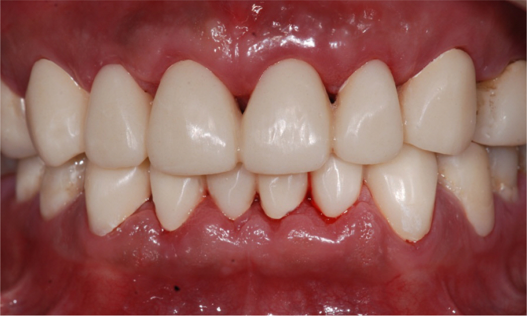
Fig. 3. Provisional prosthesis. (A) Maxillary occlusal view, (B) Lateral view (right), (C) Frontal view, (D) Lateral view (left), (E) Mandibular occlusal view.
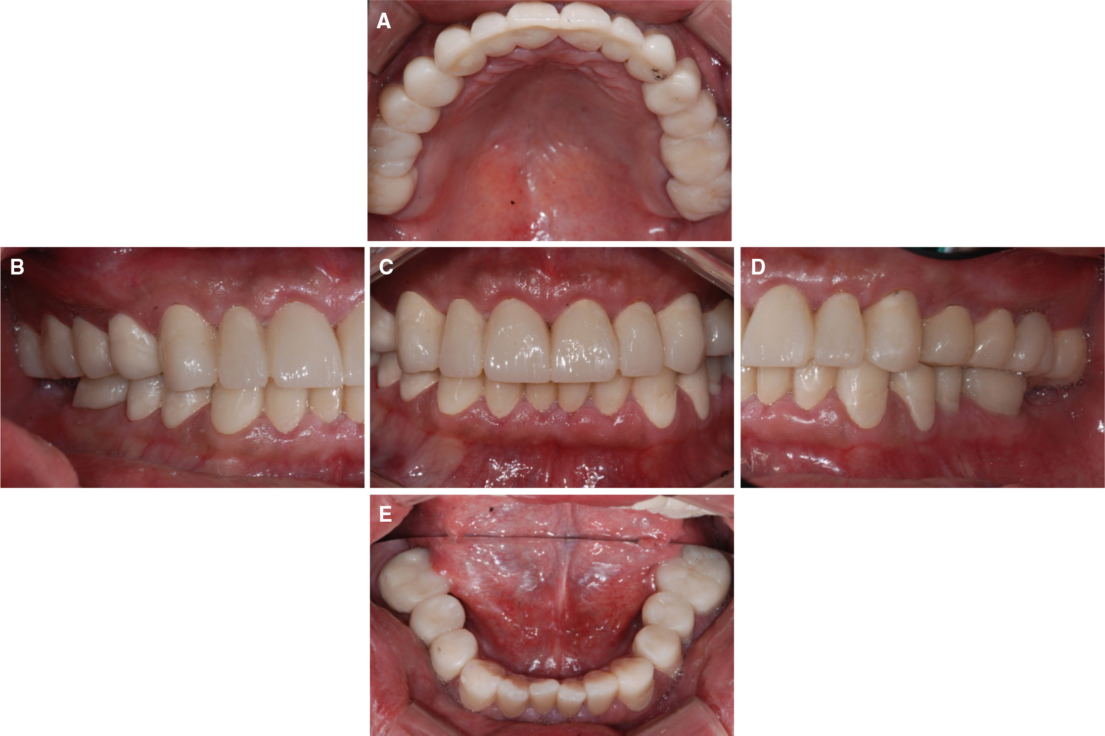
Fig. 4. Secondary provisional prosthesis.

Fig. 5. Definitive prosthesis. (A) Maxillary occlusal view, (B) Lateral view (right), (C) Frontal view, (D) Lateral view (left), (E) Right working movement, (F) Mandibular occlusal view, (G) Left working movement.
Reference
-
1.Verrett RG. Analyzing the etiology of an extremely worn dentition. J Prosthodont. 2001. 10:224–33.
Article2.Turner KA., Missirlian DM. Restoration of the extremely worn dentition. J Prosthet Dent. 1984. 52:467–74.
Article3.Briggs P., Bishop K. Fixed prostheses in the treatment of tooth wear. Eur J Prosthodont Restor Dent. 1997. 5:175–80.4.Hemmings KW., Darbar UR., Vaughan S. Tooth wear treated with direct composite restorations at an increased vertical dimension: results at 30 months. J Prosthet Dent. 2000. 83:287–93.
Article5.Berry DC., Poole DF. Attrition: possible mechanisms of compensation. J Oral Rehabil. 1976. 3:201–6.
Article6.Ramfjord SP., Blankenship JR. Increased occlusal vertical dimension in adult monkeys. J Prosthet Dent. 1981. 45:74–83.
Article7.Dawson PE. Functional occlusion: from TMJ to smile design. St. Louis; Mo: Mosby;2007. p. 430–52.8.Verrett RG. Analyzing the etiology of an extremely worn dentition. J Prosthodont. 2001. 10:224–33.
Article9.Abduo J., Lyons K. Clinical considerations for increasing occlusal vertical dimension: a review. Aust Dent J. 2012. 57:2–10.
Article
- Full Text Links
-
- Actions
-
Cited
- CITED
-
- Close
- Share
-
- Similar articles
-
- Full mouth rehabilitation using zirconia crown in severe worn dentition: a case report
- Full mouth rehabilitation of the patient with severely worn dentition and limited vertical dimension
- Oral rehabilitation of a patient with severely worn dentition using monolithic zirconia
- Full mouth rehabilitation using monolithic zirconia: a clinical report
- Full mouth rehabilitation of the patient with worn dentition using full-contour monolithic zirconia prostheses at an increased vertical dimension of occlusion: a case report
