Orthognathic surgery on Skeletal Class III patiens with collapsed vertical dimension: case report
- Affiliations
-
- 1Department of Orthodontics, Pusan National University Hospital, Busan, Republic of Korea. youngyng@hanmail.net
- 2Department of Oral and maxillofacial surgery, School of Dentistry, Pusan National University, Yangsan, Republic of Korea.
- 3Department of Orthodontics, School of Dentistry, Pusan National University, Yangsan, Republic of Korea.
- KMID: 2162381
- DOI: http://doi.org/10.14368/jdras.2016.32.1.70
Abstract
- Patients who lost posterior teeth due to periodontitis or dental caries have collapsed vertical dimension, unstable occlusion and change of the mandibular position. In particular, patients in orthognathic surgery, clinician should re-establish the pre-operative stable position of mandibular condyle in articular fossa and favorable vertical dimension for high post-operative stability of mandible. Therefore, interdisciplinary approach and co-operation, including prosthetics, orthodontics, oral and maxillofacial surgeon, from diagnosis and treatment plan is important to get a good outcome. This case report was patients who had collapsed occlusal plane due to severe dental caries on maxillary molars with skeletal Class III malocclusion. Before orthognathic surgery, resetting of maxillary occlusal plane with temporary removable denture was performed. Then successful multidisciplinary approach was done and lead to acceptable clinical outcome.
Keyword
MeSH Terms
Figure
-

Fig. 1 (A) Pre-treatment panoramic radiograph, (B) Postendodontic treatment(before orthognathic surgery) panoramic radiograph. #14, 25, 26, 36, 37, 46 were extracted.

Fig. 2 Pretreatment (before orthognathic surgery) extra (A) and intra (B) oral photos.

Fig. 3 Pretreatment (before orthognathic surgery) lateral cephalometric radiograph and analysis. *, **, *** Indicates that the value is beyond one, two or three times of the standard deviation. SNA, the angle of sella-nasion-A point; SNB, the angle of sella-nasion-B point; ANB, the angle of A point-nasion-B point; 1-SN, the angle of the long axis of maxillary central incisors between sella-nasion plane; 1-Mx.OP., the angle of the long axis of maxillary central incisors between maxillary occlusal plane; IMPA, the angle of the lowermost tangent to the mandible and the long axis of mandibular incisors; FMA, Frankfort mandibular plane angle; FH-Occ., the angle of Frankfort Horizontal plane and bisecting occlusal plane; Wits, Wits appraisal.

Fig. 4 A diagram for multidisciplinary treatment plan.

Fig. 5 Comparison of lateral cephalometric radiograph between pretreatment (A) and after maxillary temporary denture delivery (B).
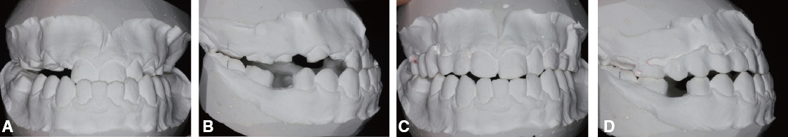
Fig. 6 Comparison cast model between pretreatment (A, B) and after maxillary temporary denture delivery (C, D).
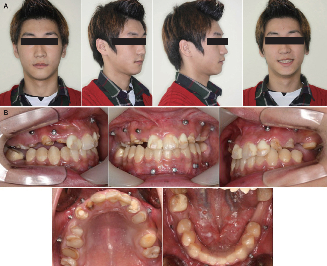
Fig. 7 Post-Orthognathic surgery (1 month) extra (A) & intra (B) oral photos. Mini-screws were inserted on both maxillary and mandibular dentoalveolar area.
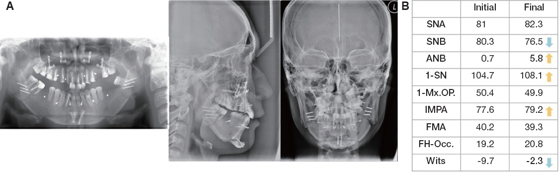
Fig. 8 Post-Orthognathic surgery (1 month) Panoramic, lateral, postero-anterior cephalometric radiograph (A) and lateral cephalometric analysis (B). SNA, the angle of sella-nasion-A point; SNB, the angle of sella-nasion-B point; ANB, the angle of A point-nasion-B point; 1-SN, the angle of the long axis of maxillary central incisors between sella-nasion plane; 1-Mx.OP., the angle of the long axis of maxillary central incisors between maxillary occlusal plane; IMPA, the angle of the lowermost tangent to the mandible and the long axis of mandibular incisors; FMA, Frankfort mandibular plane angle; FH-Occ., the angle of Frankfort Horizontal plane and bisecting occlusal plane; Wits, Wits appraisal.

Fig. 9 Set-up model for final prosthetics.

Fig. 10 Post-orthodontic treatment. Maintain vertical position on mandibular teeth. #46 was protracted.
Reference
-
References
1. Wolford LM, Chemello PD, Hilliard F. Occlusal plane alteration in orthognathic surgery – Part I effects on function and esthetics. Am J Orthod Dentofacial Orthop. 1994; 106:304–16. DOI: 10.1016/S0889-5406(94)70051-6.2. Ricketts RM. Cephalometric analysis synthesis. Angle Orthod. 1961; 31:141–56.3. Selwyn SL. An assessment of patients with periodontally involved migrated incisors. J Dent. 1973; 1:153–7. DOI: 10.1016/0300-5712(73)90027-4.4. Brunsvold MA. Pathologic tooth migration. J Periodontol. 2005; 76:859–66. DOI: 10.1902/jop.2005.76.6.859. PMID: 15948679.5. Sierpinska T, Golebiewska M, Kuc J, Lapuc M. The influence of the occlusal vertical dimension on masticatory muscle activities and hyoid bone position in complete denture wearers. Adv Med Sci. 2009; 54:104–8. DOI: 10.2478/v10039-009-0018-3. PMID: 19505871.6. Granados JI. The influence of the loss of teeth and attrition on the articular eminence. J Prosthet Dent. 1979; 42:78–85. DOI: 10.1016/0022-3913(79)90333-0.7. Uribe F, Janakiraman N, Nanda R. Interdisciplinary approach for increasing the vertical dimension of occlusion in an adult patient with several missing teeth. Am J Orthod Dentofacial Orthop. 2013; 143:867–76. DOI: 10.1016/j.ajodo.2012.05.022. PMID: 23726337.8. Abduo J. Safety of increasing vertical dimension of occlusion: a systematic review. Quintessence Int. 2012; 43:369–80. PMID: 22536588.9. Proffit WR, Bell WH. Surgical correction of dentofacial deformities. Philadelphia;WB Saunders;. 1980; 1111–3.10. Igarashi Y, Yamashita S, Kuroiwa A. Changes in interarch distance and condylar position related to loss of occlusal support for partially edentulous patients. A pilot study. Eur J Prosthodont Restor Dent. 1999; 7:107–11. PMID: 11314422.11. Willis FM. Features of the face involved in full denture prosthesis. Dent Cosmos. 1935; 77:851–4.12. Fayz F, Eslami A. Determination of occlusal vertical dimension: a literature review. J Prosthet Dent. 1988; 59:321–3. DOI: 10.1016/0022-3913(88)90182-5.13. Spalding PM, Cohen BD. Orthodontic adjunctive treatment in fixed prosthodontics. Dent Clin North Am. 1992; 36:607–29. PMID: 1397428.14. Miller TE. Orthodontic therapy for the restorative patient. Part I: the biomechanic aspects. J Prosthet Dent. 1989; 61:268–76. DOI: 10.1016/0022-3913(89)90126-1.15. Hwang HS, Lee KH. Intrusion of overerupted molars by corticotomy and magnets. Am J Orthod Dentofacial Orthop. 2001; 120:209–16. DOI: 10.1067/mod.2001.115149. PMID: 11500664.16. Mostafa YA, Tawfik KM, El-Mangoury NH. Surgical- orthodontic treatment for overerupted maxillary molars. J Clin Orthod. 1985; 19:350–1. PMID: 3859489.17. Park YC, Lee SY, Kim DH, Jee SH. Intrusion of posterior teeth using mini-screw implants. Am J Orthod Dentofacial Orthop. 2003; 123:690–4. DOI: 10.1016/S0889-5406(03)00047-7.18. Dellinger EL. A histologic and cephalometric investigation of premolar intrusion in the Macaca speciosa monkey. Am J Orthod. 1967; 53:325–55. DOI: 10.1016/0002-9416(67)90100-5.19. Kalra V, Burstone CJ, Nanda R. Effects of a fixed magnetic appliance on the dentofacial complex. Am J Orthod Dentofacial Orthop. 1989; 95:467–78. DOI: 10.1016/0889-5406(89)90410-1.
- Full Text Links
-
- Actions
-
Cited
- CITED
-
- Close
- Share
-
- Similar articles
-
- Two treatment approach to skeletal Class III : A case report on sisters
- Multidisciplinary management for amelogenesis imperfecta patient with skeletal CIII malocclusion
- Full-mouth rehabilitation with increasing vertical dimension on the patient with severely worn-out dentition and orthognathic surgery history: A case report
- The study of the soft tissue change according to skeletal change following bssro with advancing genioplasty
- Changes of the hyoid bone position and the upper airway dimension after orthognathic surgery in skeletal class III patients
