Differential Imaging Features of Pulmonary Artery Dissection from Other Intraluminal Diseases of Pulmonary Artery: Two Cases Report
- Affiliations
-
- 1Department of Radiology, Daegu Fatima Hospital, Daegu, Korea. hwshin@fatima.or.kr
- 2Department of Pathology, Daegu Fatima Hospital, Daegu, Korea.
- KMID: 2161173
- DOI: http://doi.org/10.3348/jksr.2015.72.3.193
Abstract
- Pulmonary artery dissection is rarer than other intraluminal diseases of pulmonary artery such as pulmonary thromboembolism or pulmonary artery sarcoma. We report two cases of pulmonary artery dissection mimicking pulmonary artery sarcoma. Computed tomography (CT) showed no enhancement of intrapulmonary arterial lesion or expansion of involved pulmonary artery. Magnetic resonance imaging (MRI) showed low-signal intensity intimal flap on T1- and T2-weighted images. There was no fluorodeoxyglucose (FDG) uptake on positron emission tomography (PET)-CT. In this case report, we describe the imaging features of pulmonary artery dissection on CT, MRI, and PET-CT.
MeSH Terms
Figure
-

Fig. 1 Contrast-enhanced axial CT (A) shows homogenous low density lesion (star) in left main pulmonary artery that occupies the entire lumen of the pulmonary artery. T1- (B) and T2- (C) weighted images show a relatively homogeneous high signal intensity lesion (star), covered by low-signal intensity line (arrow) (case 1).

Fig. 2 Non-enhanced axial CT (A) and contrast-enhanced axial CT (B) show no enhancement of left main pulmonary arterial lesion (star). Contrast enhanced coronal CT (C) shows that the lesion (star) extends to left lower lobar artery (arrow). Positron emission tomography-CT (D) shows negative fluorodeoxyglucose (FDG) uptake (star). Calcified right perihilar lymph nodes (open arrow) show mild FDG uptake (case 2).
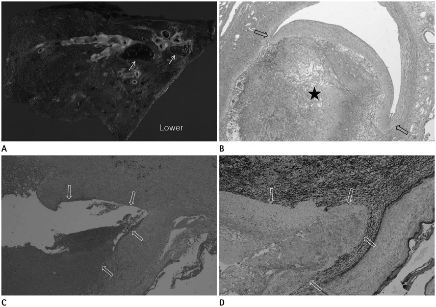
Fig. 3 68-year-old woman patient's gross specimen (A) shows hematoma in the pulmonary artery (arrows). Microscopic examination (hematoxylin and eosin stain, × 20) (B) reveals that the hematoma (star) is located in dissected smooth muscle layer (open arrows). 48-year-old woman patient's hematoxylin and eosin stain (× 20) (C) and elastic stain (× 20) (D) of specimen show that the smooth muscle of the pulmonary artery split into two layers (arrows). A, B. Case 2. C, D. Case 1.
Reference
-
1. Mohammad K, Sahlol M, Egiebor O, Sadikot RT. Idiopathic pulmonary artery dissection: a case report. J Med Case Rep. 2009; 3:7426.2. Neimatallah MA, Hassan W, Moursi M, Al Kadhi Y. CT findings of pulmonary artery dissection. Br J Radiol. 2007; 80:e61–e63.3. Stern EJ, Graham C, Gamsu G, Golden JA, Higgins CB. Pulmonary artery dissection: MR findings. J Comput Assist Tomogr. 1992; 16:481–483.4. Degano B, Prevot G, Têtu L, Sitbon O, Simonneau G, Humbert M. Fatal dissection of the pulmonary artery in pulmonary arterial hypertension. Eur Respir Rev. 2009; 18:181–185.5. Wunderbaldinger P, Bernhard C, Uffmann M, Kürkciyan I, Senbaklavaci O, Herold CJ. Acute pulmonary trunk dissection in a patient with primary pulmonary hypertension. J Comput Assist Tomogr. 2000; 24:92–95.6. Allkemper T, Tombach B, Schwindt W, Kugel H, Schilling M, Debus O, et al. Acute and subacute intracerebral hemorrhages: comparison of MR imaging at 1.5 and 3.0 T--initial experience. Radiology. 2004; 232:874–881.7. Bressler EL, Nelson JM. Primary pulmonary artery sarcoma: diagnosis with CT, MR imaging, and transthoracic needle biopsy. AJR Am J Roentgenol. 1992; 159:702–704.8. Yi CA, Lee KS, Choe YH, Han D, Kwon OJ, Kim S. Computed tomography in pulmonary artery sarcoma: distinguishing features from pulmonary embolic disease. J Comput Assist Tomogr. 2004; 28:34–39.9. Chong S, Kim TS, Kim BT, Cho EY, Kim J. Pulmonary artery sarcoma mimicking pulmonary thromboembolism: integrated FDG PET/CT. AJR Am J Roentgenol. 2007; 188:1691–1693.10. Thurer RL, Thorsen A, Parker JA, Karp DD. FDG imaging of a pulmonary artery sarcoma. Ann Thorac Surg. 2000; 70:1414–1415.
- Full Text Links
-
- Actions
-
Cited
- CITED
-
- Close
- Share
-
- Similar articles
-
- Aortic Dissection Presenting with Secondary Pulmonary Hypertension Caused by Compression of the Pulmonary Artery by Dissecting Hematoma: A Case Report
- Pulmonary artery sling: case report
- Spontaneous Resolution of Pulmonary Artery Pseudoaneurysm after Tube Thoracostomy
- Pulmonary Artery Intimal Sarcoma Involving the Peripheral Pulmonary Artery, Initially Misdiagnosed as Pulmonary Artery Thromboembolism and Vasculitis: A Case Report
- RVOTO Caused by Pulmonary Artery Sarcoma Originating from Pulmonary Valve: One case report
