Stormy Course of a Huge Submitral Aneurysm Causing Low Cardiac Output State
- Affiliations
-
- 1Post Graduate Department of Cardiology, JLN Medical College & Associated Group of Hospitals, Ajmer, India. avinboxer@gmail.com
- KMID: 2160999
- DOI: http://doi.org/10.4250/jcu.2016.24.1.68
Abstract
- Submitral aneurysm is a rare structural abnormality of congenital or acquired aetiology. Most reported cases are from Africa. Unless promptly treated surgically this condition is invariably fatal. We report a case of a young Indian male who presented with dyspnea of recent onset, diagnosed to have a massive submitral aneurysm causing low cardiac output and compression of cardiac structures.
Keyword
Figure
-

Fig. 1 Two dimensional echocardiographic images acquired using Philips iE33/S5-1 probe. A: Apical 4 chamber view showing a large cystic mass of size comparable to both the ventricles put together. B: Same view with plane shifted anteriorly to visualize the communication (*) between the cyst and the left ventricle (just beneath the posterior mitral leaflet). C and D: Parasternal short axis view at the basal level (with and without color Doppler, respectively), showing the giant aneurysm compressing left ventricle, with bidirectional flow between the two, largest dimension of the aneurysm being 7.46 × 8.53 cm. AML: anterior mitral leaflet, PML: posterior mitral leaflet, PW: posterior wall of LV, IVS: inter ventricular septum, Ao: aorta, LV: left ventricle, RV: right ventricle.
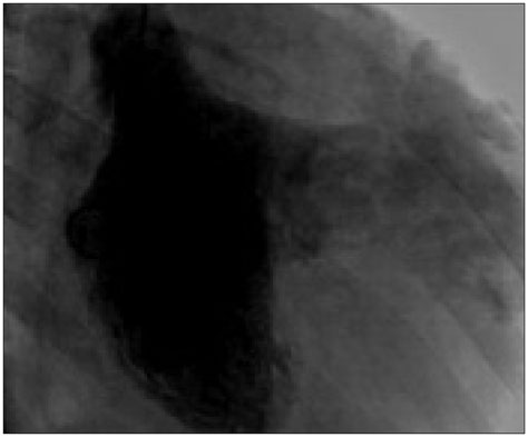
Fig. 2 Left ventricular angiogram in right anterior oblique view with 30° angulation showing the giant aneurysm in free communication with the left ventricle at the basal level, its huge dimensions completely displacing the left ventricle from its original site. Pig tail catheter in situ seen.
Reference
-
1. Chesler E, Joffe N, Schamroth L, Meyers A. Annular subvalvular left ventricular aneurysms in the South African Bantu. Circulation. 1965; 32:43–51.2. Abrahams DG, Barton CJ, Cockshott WP, Edington GM, Weaver EJ. Annular subvalvular left ventricular aneurysms. Q J Med. 1962; 31:345–360.3. Chugh VK, Sabharwal U. Subvalvular cardiac aneurysm. Indian Heart J. 1978; 30:171–173.4. Ribeiro PJ, Mendes RG, Vicente WV, Menardi AC, Evora PR. Submitral left ventricular aneurysm. Case report and review of published Brazilian cases. Arq Bras Cardiol. 2001; 76:395–402.5. Wolpowitz A, Arman B, Barnard MS, Barnard CN. Annular subvalvular idiopathic left ventricular aneurysms in the black African. Ann Thorac Surg. 1979; 27:350–355.6. Chockalingam A, Gnanavelu G, Alagesan R, Subramaniam T. Congenital submitral aneurysm and sinus of valsalva aneurysm. Echocardiography. 2004; 21:325–328.7. Cavallé-Garrido T, Cloutier A, Harder J, Boutin C, Smallhorn JF. Evolution of fetal ventricular aneurysms and diverticula of the heart: an echocardiographic study. Am J Perinatol. 1997; 14:393–400.8. Awasthy N, Shrivastava S. Submitral aneurysm: an antenatal diagnosis. Ann Pediatr Cardiol. 2013; 6:164–166.9. Esposito F, Renzulli A, Festa M, Cerasuolo F, Caruso A, Sarnicola P, Cotrufo M. Submitral left ventricular aneurysm. Report of 2 surgical cases. Tex Heart Inst J. 1996; 23:51–53.10. Mohan JC, Goel PK, Khanna SK, Arora R. Massive congenital submitral aneurysm of the left ventricle: a case report. Indian Heart J. 1989; 41:338–340.
- Full Text Links
-
- Actions
-
Cited
- CITED
-
- Close
- Share
-
- Similar articles
-
- Severe Vertebral Erosion by Huge Symptomatic Pulsating Aortic Aneurysm
- Surgical Treatment of a Submitral Left Ventricular Aneurysm and the Patient Present with Recurrent Ventricular Tachycardia
- The Cardiac Output and the Cardiac Muscle Contractility During Postural Gradient Changes by Tilt Table in Man
- Surgical Management of an Isolated Huge Innominate Artery Aneurysm Causing Tracheal Compression: A Case Report
- Comparison of Left and Right Ventricular Volume and Cardiac Output by MRI and Echocardiography
