Ann Surg Treat Res.
2016 Apr;90(4):231-234. 10.4174/astr.2016.90.4.231.
Unusual intestinal obstruction due to idiopathic sclerosing encapsulating peritonitis: a report of two cases and a review
- Affiliations
-
- 1Department of Surgery, Daegu Catholic University Medical Center, Catholic University of Daegu School of Medicine, Daegu, Korea. silverpop@hanmail.net
- KMID: 2160954
- DOI: http://doi.org/10.4174/astr.2016.90.4.231
Abstract
- Sclerosing encapsulating peritonitis (SEP) is a rare cause of intestinal obstruction that is characterized by a thick fibrotic membrane encasing the small intestine like a cocoon. Accurate preoperative diagnosis is often difficult. We present 2 cases of SEP that were diagnosed preoperatively by contrast-enhanced computed tomography scan. A 38-year-old man and a 56-year-old woman were admitted to Daegu Catholic University Medical Center because of recurrent intestinal obstruction. We performed exploratory laparotomy with doubt of the preoperative diagnosis of SEP. We confirmed the diagnosis of SEP on laparotomy and performed adhesiolysis. Both patients recovered successfully and had no signs of recurrence. A better awareness of SEP and its radiological features should lead to more correct preoperative diagnosis and result in more appropriate management, including surgery.
MeSH Terms
Figure
-

Fig. 1 The axial section shows marked dilatation and thickening of the small intestine, encased in a thick enhancing membrane. Fluid collection between the bowel loops is seen.
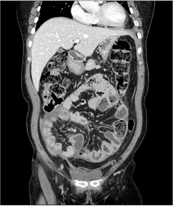
Fig. 2 The coronal section reveals congregated loops to the center of the abdomen by the thickened peritoneum.
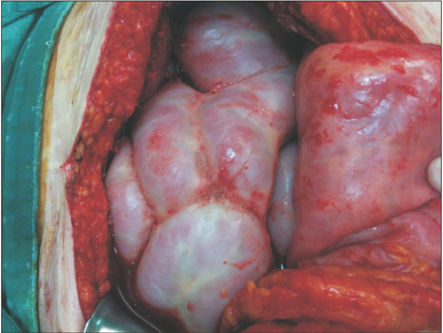
Fig. 3 At laparotomy, the small intestine is encased in a cocoon-like fibrotic tissue.
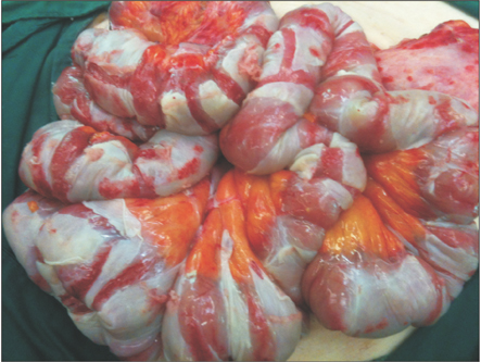
Fig. 4 After peritoneal decortication, we can see the fibrous membranes.
Reference
-
1. Foo KT, Ng KC, Rauff A, Foong WC, Sinniah R. Unusual small intestinal obstruction in adolescent girls: the abdominal cocoon. Br J Surg. 1978; 65:427–430.2. Akbulut S. Accurate definition and management of idiopathic sclerosing encapsulating peritonitis. World J Gastroenterol. 2015; 21:675–687.3. Tannoury JN, Abboud BN. Idiopathic sclerosing encapsulating peritonitis: abdominal cocoon. World J Gastroenterol. 2012; 18:1999–2004.4. Wei B, Wei HB, Guo WP, Zheng ZH, Huang Y, Hu BG, et al. Diagnosis and treatment of abdominal cocoon: a report of 24 cases. Am J Surg. 2009; 198:348–353.5. Xu P, Chen LH, Li YM. Idiopathic sclerosing encapsulating peritonitis (or abdominal cocoon): a report of 5 cases. World J Gastroenterol. 2007; 13:3649–3651.6. Devay AO, Gomceli I, Korukluoglu B, Kusdemir A. An unusual and difficult diagnosis of intestinal obstruction: The abdominal cocoon. Case report and review of the literature. World J Emerg Surg. 2008; 3:36.7. George C, Al-Zwae K, Nair S, Cast JE. Computed tomography appearances of sclerosing encapsulating peritonitis. Clin Radiol. 2007; 62:732–737.8. Honda K, Oda H. Pathology of encapsulating peritoneal sclerosis. Perit Dial Int. 2005; 25:Suppl 4. S19–S29.9. Singh B, Gupta S. Abdominal cocoon: a case series. Int J Surg. 2013; 11:325–328.10. Habib SM, Betjes MG, Fieren MW, Boeschoten EW, Abrahams AC, Boer WH, et al. Management of encapsulating peritoneal sclerosis: a guideline on optimal and uniform treatment. Neth J Med. 2011; 69:500–507.
- Full Text Links
-
- Actions
-
Cited
- CITED
-
- Close
- Share
-
- Similar articles
-
- Sclerosing Encapsulating Peritonitis in Chronic Ambulatory Peritoneal Dialysis; Preoperative Catheter Drainage: A Case Report
- Sclerosing encapsulating peritonitis after living-donor liver transplantation: A case series, Kyoto experience
- Sclerosing Encapsulating Peritonitis (Abdominal Cocoon) after Abdominal Hysterectomy
- Ultrasonographic Findings of Sclerosing Encapsulating Peritonitis
- Successful Treatment of Sclerosing Encapsulating Peritonitis with Tamoxifen and Prednisolone
