Primary Pulmonary Low-Grade Angiosarcoma Characterized by Mismatch between 18F-FDG PET and Dynamic Contrast-Enhanced CT
- Affiliations
-
- 1Department of Radiology and Center for Imaging Science, Samsung Medical Center, Sungkyunkwan University School of Medicine, Seoul 06351, Korea. hoyunlee96@gmail.com
- 2Department of Pathology, Samsung Medical Center, Sungkyunkwan University School of Medicine, Seoul 06351, Korea.
- 3Department of Nuclear Medicine, Samsung Medical Center, Sungkyunkwan University School of Medicine, Seoul 06351, Korea.
- KMID: 2160785
- DOI: http://doi.org/10.3348/kjr.2015.16.5.1166
Abstract
- We report a rare case of primary pulmonary low-grade angiosarcoma on dynamic contrast-enhanced CT and 18F-fluorodeoxyglucose (FDG) positron emission tomography (PET)/CT imaging. A 38-year-old, asymptomatic woman was hospitalized because of an abnormality on chest radiography. A dynamic contrast-enhanced chest CT showed a 1.2 cm-sized irregular-margined nodule with strong and persistent enhancement in the right lower lobe. The lesion had low metabolic activity on an 18F-FDG PET/CT scan. The patient underwent a wedge resection for the lesion, and pathology revealed a primary pulmonary low-grade angiosarcoma.
MeSH Terms
-
Adult
Female
Fluorodeoxyglucose F18/*chemistry
Hemangiosarcoma/*diagnosis/pathology/radiography
Humans
Ki-67 Antigen/metabolism
Lung Neoplasms/*diagnosis/pathology/radiography
Multimodal Imaging
*Positron-Emission Tomography
Radiopharmaceuticals/*chemistry
Tomography, Spiral Computed
Fluorodeoxyglucose F18
Ki-67 Antigen
Radiopharmaceuticals
Figure
-
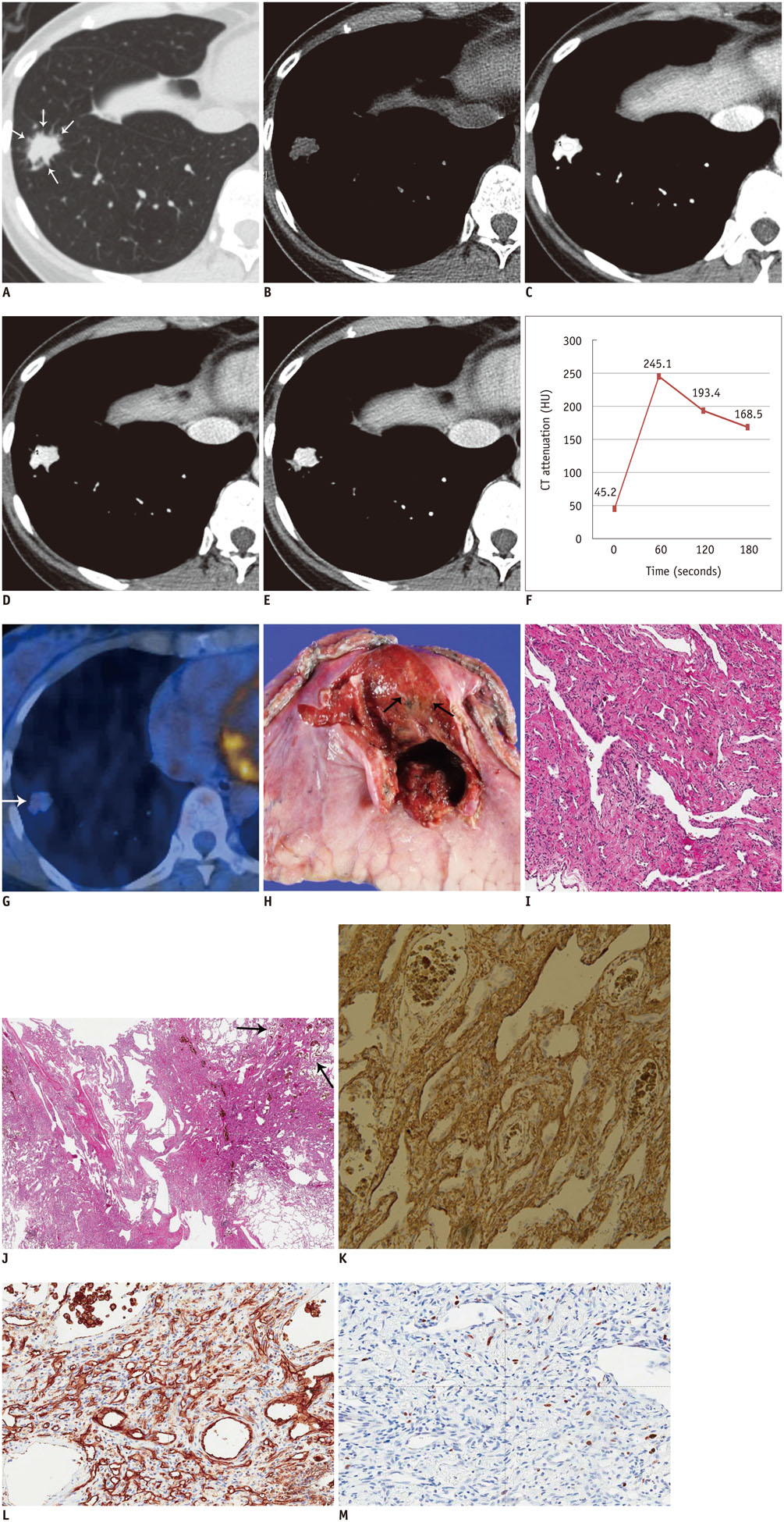
Fig. 1 38-year-old woman with primary pulmonary angiosarcoma. A. Transverse lung-window (window level of -700 H and window width of 1500 H) CT image indicates 1.2 cm-sized nodule in right lower lobe with spiculated margin and needle-like projections (arrows) in periphery of nodule. B-F. Serial transverse images obtained through nodule for 180 seconds allowed dynamic enhancement curve for nodule to be plotted. Graph shows early peak enhancement and gradual loss of enhancement (washout). G. 18F-fluorodeoxyglucose positron emission tomography fused axial image shows nodule with mildly increased radiopharmaceutical uptake (arrow) with maximum standardized uptake value of 2.0. H. Gross photograph indicates soft and dark-reddish nodule with hemorrhage (arrows). I. Photomicrograph reveals highly vascular tumor with prominent freely anastomosing vascular channels, papillary growth, and endothelial tufting that was absent to minimal (× 200). J. Tumor's peripheral vasoformative features (arrows) indicate anastomosing vascular channels lined by malignant endothelium (× 100). These marginal characteristics correspond to needle-like projections in periphery of nodule seen in CT image A. K-M. Immunohistochemistry staining shows tumor cells are diffusely positive for SMA (K), and CD31 (L), and weakly positive for Ki-67 (M) (× 200). SMA = smooth muscle actin
Reference
-
1. Chen YB, Guo LC, Yang L, Feng W, Zhang XQ, Ling CH, et al. Angiosarcoma of the lung: 2 cases report and literature reviewed. Lung Cancer. 2010; 70:352–356.2. Eichner R, Schwendy S, Liebl F, Huber A, Langer R. Two cases of primary pulmonary angiosarcoma as a rare cause of lung haemorrhage. Pathology. 2011; 43:386–389.3. Patel AM, Ryu JH. Angiosarcoma in the lung. Chest. 1993; 103:1531–1535.4. Weissferdt A, Moran CA. Primary vascular tumors of the lungs: a review. Ann Diagn Pathol. 2010; 14:296–308.5. Jeong YJ, Lee KS, Jeong SY, Chung MJ, Shim SS, Kim H, et al. Solitary pulmonary nodule: characterization with combined wash-in and washout features at dynamic multi-detector row CT. Radiology. 2005; 237:675–683.6. Young RJ, Brown NJ, Reed MW, Hughes D, Woll PJ. Angiosarcoma. Lancet Oncol. 2010; 11:983–991.7. Bastiaannet E, Groen H, Jager PL, Cobben DC, van der Graaf WT, Vaalburg W, et al. The value of FDG-PET in the detection, grading and response to therapy of soft tissue and bone sarcomas; a systematic review and meta-analysis. Cancer Treat Rev. 2004; 30:83–101.8. Eary JF, Conrad EU, Bruckner JD, Folpe A, Hunt KJ, Mankoff DA, et al. Quantitative [F-18] fluorodeoxyglucose positron emission tomography in pretreatment and grading of sarcoma. Clin Cancer Res. 1998; 4:1215–1220.9. Tokmak E, Ozkan E, Yağcı S, Kır KM. F18-FDG PET/CT Scanning in Angiosarcoma: Report of Two Cases. Mol Imaging Radionucl Ther. 2011; 20:63–66.10. Cucci E, Ciuffreda M, Tambaro R, Aquilani L, Barrassi M, Sallustio G. MRI findings of large low-grade angiosarcoma of the breast with subsequent bone metastases: a case report. J Breast Cancer. 2012; 15:255–257.11. Lim RF, Goei R. Best cases from the AFIP: angiosarcoma of the breast. Radiographics. 2007; 27:Suppl 1. S125–S130.
- Full Text Links
-
- Actions
-
Cited
- CITED
-
- Close
- Share
-
- Similar articles
-
- Supraclavicular Lymph Node Metastasis from Various Malignancies: Assessment with 18F-Fluorodeoxyglucose Positron Emission Tomography/CT, Contrast-Enhanced CT and Ultrasound
- Use of 18F-FDG PET/CT in Second Primary Cancer
- Uterine Epithelioid Angiosarcoma on F-18 FDG PET/CT
- Usefulness of Low Dose Oral Contrast Media in 18F-FDG PET/CT
- Incidental Bilateral Renal Oncocytoma in a Patient with Metastatic Carcinoma of Unknown Primary: a Pitfall on 18F-FDG PET/CT
