Treatment Response Evaluation of Breast Cancer after Neoadjuvant Chemotherapy and Usefulness of the Imaging Parameters of MRI and PET/CT
- Affiliations
-
- 1Department of Radiology, College of Medicine, The Catholic University of Korea, St. Vincent's Hospital, Suwon, Korea.
- 2Department of Radiology, College of Medicine, The Catholic University of Korea, Seoul St. Mary's Hospital, Seoul, Korea.
- 3Department of Hospital Pathology, College of Medicine, The Catholic University of Korea, Seoul St. Mary's Hospital, Seoul, Korea. didi97@catholic.ac.kr
- KMID: 2160612
- DOI: http://doi.org/10.3346/jkms.2015.30.6.808
Abstract
- This study was aimed to evaluate the ability of imaging parameters measured on dynamic contrast-enhanced magnetic resonance imaging (DCE-MRI), diffusion-weighted MRI (DWI) and positron emission tomography/computed tomography (PET/CT) to serve as response markers in breast cancer after neoadjuvant chemotherapy (NAC). In 20 patients with breast cancer, DCE-MRI and DWI using a 3 T scanner and PET/CT were performed before and after NAC. DCE-MRI was analyzed using an automatic computer-aided detection program (MR-CAD). The response imaging parameters were compared with the pathologic response. The areas under the curve (AUCs) for DCE-MRI using MR-CAD analysis, DWI and PET/CT were 0.77, 0.59 and 0.76, respectively. The combination of all parameters measured by MR-CAD showed the highest diagnostic performance and accuracy (AUC = 0.77, accuracy = 90%). The combined use of the parameters of PET/CT with DCE-MRI or DWI showed a trend toward improved specificity and negative predictive value (100%, 100%, accuracy = 87.5%). The use of DCE-MRI using MR-CAD parameters indicated better diagnostic performance in predicting the final pathological response compared with DWI and PET/CT, although no statistically significant difference was observed. The combined use of PET/CT with DCE-MRI or DWI may improve the specificity for predicting a pathological response.
Keyword
MeSH Terms
-
Adult
Aged
Antineoplastic Agents/*therapeutic use
Breast Neoplasms/*diagnosis/*drug therapy
Chemotherapy, Adjuvant/methods
Female
Humans
Image Interpretation, Computer-Assisted/methods
Magnetic Resonance Imaging/*methods
Mammography/methods
Middle Aged
Multimodal Imaging/methods
Neoadjuvant Therapy/methods
Positron-Emission Tomography/*methods
Reproducibility of Results
Retrospective Studies
Sensitivity and Specificity
Tomography, X-Ray Computed/*methods
Treatment Outcome
Antineoplastic Agents
Figure
-

Fig. 1 Examples of the response evaluation using MR-CAD, DWI and PET/CT. (A) DCE-MRI analysis using MR-CAD provided information regarding the size, volume and kinetics of a tumor using automatic segmentation. (B) For DWI analysis, the ADC values were obtained by manually drawing an ROI within a hypointense tumor on the ADC map. (C) For PET/CT analysis, ROIs were manually placed over tumors in attenuation-corrected images, and the peak standardized uptake values (pSUV) within the ROIs were recorded.
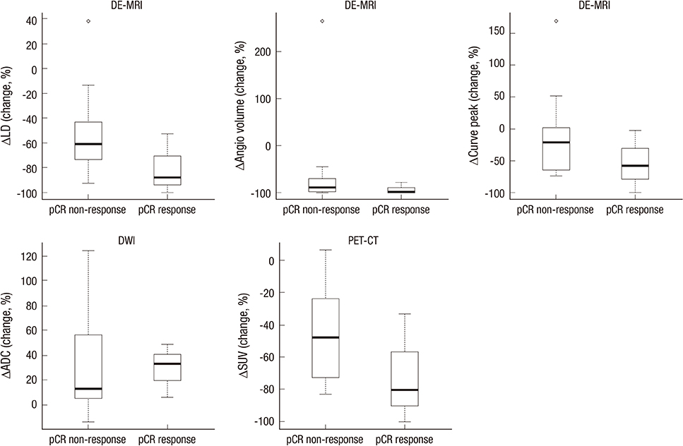
Fig. 2 Box-plots comparing the percent changes of the quantitative parameters in DCE-MRI (LD, TV, PE), DWI (ADC) and PET/CT (SUV max). The mean percent change of the imaging parameters measured by DCE-MRI and PET/CT was decreased more in responders than in non-responders. By contrast, the mean percent change of the ADC value was increased more in the responder group.
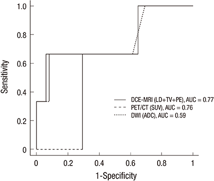
Fig. 3 ROC curve analyses of DCE-MRI, DWI and PET/CT for the prediction of pathologic responses. The AUC values for the DCE-MRI, DWI and PET/CT were 0.77 (95% CI = 0.28 to 0.89), 0.59 (95% CI = 0.28 to 0.89) and 0.76 (95% CI = 0.34 to 1.00), respectively. The DCE-MRI analysis using all three CAD parameters resulted in the highest diagnostic performance compared with DWI or PET/CT.
Reference
-
1. Mauri D, Pavlidis N, Ioannidis JP. Neoadjuvant versus adjuvant systemic treatment in breast cancer: a meta-analysis. J Natl Cancer Inst. 2005; 97:188–194.2. Kong X, Moran MS, Zhang N, Haffty B, Yang Q. Meta-analysis confirms achieving pathological complete response after neoadjuvant chemotherapy predicts favourable prognosis for breast cancer patients. Eur J Cancer. 2011; 47:2084–2090.3. Gonzalez-Angulo AM, Morales-Vasquez F, Hortobagyi GN. Overview of resistance to systemic therapy in patients with breast cancer. Adv Exp Med Biol. 2007; 608:1–22.4. Drew PJ, Chatterjee S, Turnbull LW, Read J, Carleton PJ, Fox JN, Monson JR, Kerin MJ. Dynamic contrast enhanced magnetic resonance imaging of the breast is superior to triple assessment for the pre-operative detection of multifocal breast cancer. Ann Surg Oncol. 1999; 6:599–603.5. Van Goethem M, Schelfout K, Kersschot E, Colpaert C, Verslegers I, Biltjes I, Tjalma WA, De Schepper A, Weyler J, Parizel PM. MR mammography is useful in the preoperative locoregional staging of breast carcinomas with extensive intraductal component. Eur J Radiol. 2007; 62:273–282.6. Eisenhauer EA, Therasse P, Bogaerts J, Schwartz LH, Sargent D, Ford R, Dancey J, Arbuck S, Gwyther S, Mooney M, et al. New response evaluation criteria in solid tumours: revised RECIST guideline (version 1.1). Eur J Cancer. 2009; 45:228–247.7. Wahl RL, Jacene H, Kasamon Y, Lodge MA. From RECIST to PERCIST: Evolving Considerations for PET response criteria in solid tumors. J Nucl Med. 2009; 50:122s–150s.8. Symmans WF, Peintinger F, Hatzis C, Rajan R, Kuerer H, Valero V, Assad L, Poniecka A, Hennessy B, Green M, et al. Measurement of residual breast cancer burden to predict survival after neoadjuvant chemotherapy. J Clin Oncol. 2007; 25:4414–4422.9. Hylton NM, Blume JD, Bernreuter WK, Pisano ED, Rosen MA, Morris EA, Weatherall PT, Lehman CD, Newstead GM, Polin S, ACRIN 6657 Trial Team and I-SPY 1 TRIAL Investigators, et al. Locally advanced breast cancer: MR imaging for prediction of response to neoadjuvant chemotherapy--results from ACRIN 6657/I-SPY TRIAL. Radiology. 2012; 263:663–672.10. Wu LM, Hu JN, Gu HY, Hua J, Chen J, Xu JR. Can diffusion-weighted MR imaging and contrast-enhanced MR imaging precisely evaluate and predict pathological response to neoadjuvant chemotherapy in patients with breast cancer? Breast Cancer Res Treat. 2012; 135:17–28.11. Lobbes MB, Prevos R, Smidt M, Tjan-Heijnen VC, van Goethem M, Schipper R, Beets-Tan RG, Wildberger JE. The role of magnetic resonance imaging in assessing residual disease and pathologic complete response in breast cancer patients receiving neoadjuvant chemotherapy: a systematic review. Insights Imaging. 2013; 4:163–175.12. Park SH, Moon WK, Cho N, Chang JM, Im SA, Park IA, Kang KW, Han W, Noh DY. Comparison of diffusion-weighted MR imaging and FDG PET/CT to predict pathological complete response to neoadjuvant chemotherapy in patients with breast cancer. Eur Radiol. 2012; 22:18–25.13. Shin HJ, Baek HM, Ahn JH, Baek S, Kim H, Cha JH, Kim HH. Prediction of pathologic response to neoadjuvant chemotherapy in patients with breast cancer using diffusion-weighted imaging and MRS. NMR Biomed. 2012; 25:1349–1359.14. Choi JH, Lim HI, Lee SK, Kim WW, Kim SM, Cho E, Ko EY, Han BK, Park YH, Ahn JS, et al. The role of PET CT to evaluate the response to neoadjuvant chemotherapy in advanced breast cancer: comparison with ultrasonography and magnetic resonance imaging. J Surg Oncol. 2010; 102:392–397.15. Dose-Schwarz J, Tiling R, Avril-Sassen S, Mahner S, Lebeau A, Weber C, Schwaiger M, Jänicke F, Untch M, Avril N. Assessment of residual tumour by FDG-PET: conventional imaging and clinical examination following primary chemotherapy of large and locally advanced breast cancer. Br J Cancer. 2010; 102:35–41.16. Park JS, Moon WK, Lyou CY, Cho N, Kang KW, Chung JK. The assessment of breast cancer response to neoadjuvant chemotherapy: comparison of magnetic resonance imaging and 18F-fluorodeoxyglucose positron emission tomography. Acta Radiol. 2011; 52:21–28.17. Tateishi U, Miyake M, Nagaoka T, Terauchi T, Kubota K, Kinoshita T, Daisaki H, Macapinlac HA. Neoadjuvant chemotherapy in breast cancer: prediction of pathologic response with PET/CT and dynamic contrast-enhanced MR imaging--prospective assessment. Radiology. 2012; 263:53–63.
- Full Text Links
-
- Actions
-
Cited
- CITED
-
- Close
- Share
-
- Similar articles
-
- Erratum: Correction of Table: Treatment Response Evaluation of Breast Cancer after Neoadjuvant Chemotherapy and Usefulness of the Imaging Parameters of MRI and PET/CT
- Response Evaluation to Neoadjuvant Chemotherapy in Advanced Breast Cancer: Comparison of MRI and Positron Emission Tomography/CT (RECIST 1.1 versus PERCIST 1.0)
- Interpretation of Rectal MRI after Neoadjuvant Treatment in Patients with Rectal Cancer
- Usefulness of Combined Metabolic-Volumetric Indices of 18F-FDG PET/CT for the Early Prediction of Neoadjuvant Chemotherapy Outcomes in Breast Cancer
- The Role of fluorodeoxyglucose PET in the management of breast cancer
