Early Retinal Changes in Hunter Syndrome According to Spectral Domain Optical Coherence Tomography
- Affiliations
-
- 1Department of Ophthalmology, Seoul National University Bundang Hospital, Seoul National University College of Medicine, Seongnam, Korea. eye@snubh.org
- KMID: 2160468
- DOI: http://doi.org/10.3341/kjo.2016.30.2.151
Abstract
- No abstract available.
Figure
-
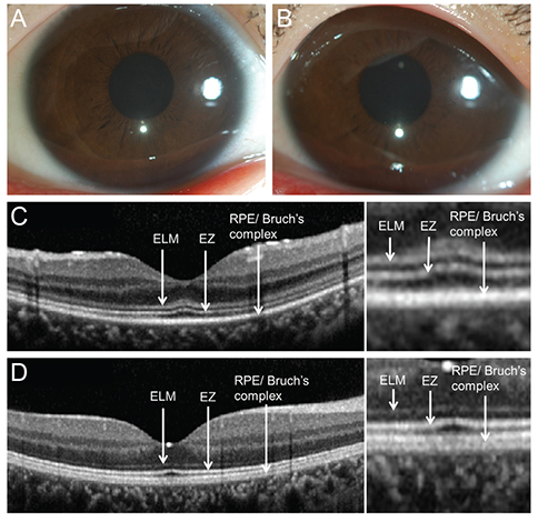
Fig. 1 Slit lamp biomicroscopy showed clear corneas in the right (A) and left (B) eyes. (C,D) Comparison of spectral domain optical coherence tomography (SD-OCT; Spectralis, Heidelberg Engineering, Heidelberg, Germany) scans of the macula in early stage Hunter syndrome and normal control. (C) Vertical scan images with SD-OCT of the patient's right eye. There was marked thickening of the external limiting membrane (ELM). The ellipsoid zone (EZ) formerly known as the inner and outer segment photoreceptor junction appeared normal in thickness and contour, well distinguished from the retinal pigment epithelial layer underneath. The photoreceptor layer and retinal pigment epithelium-Bruch's membrane complex (RPE/Bruch's complex) showed no thinning or other abnormal change. (D) SD-OCT of the normal eye from a control subject showed four distinct high-signal bands in the outer retinal layer without foveal thickening of the ELM.
Reference
-
1. Ashworth JL, Biswas S, Wraith E, Lloyd IC. Mucopolysaccharidoses and the eye. Surv Ophthalmol. 2006; 51:1–17.2. McDonnell JM, Green WR, Maumenee IH. Ocular histopathology of systemic mucopolysaccharidosis, type II-A (Hunter syndrome, severe). Ophthalmology. 1985; 92:1772–1779.3. Yoon MK, Chen RW, Hedges TR 3rd, et al. High-speed, ultrahigh resolution optical coherence tomography of the retina in Hunter syndrome. Ophthalmic Surg Lasers Imaging. 2007; 38:423–428.4. Chen TC, Cense B, Pierce MC, et al. Spectral domain optical coherence tomography: ultra-high speed, ultra-high resolution ophthalmic imaging. Arch Ophthalmol. 2005; 123:1715–1720.5. Bunt-Milam AH, Saari JC, Klock IB, Garwin GG. Zonulae adherentes pore size in the external limiting membrane of the rabbit retina. Invest Ophthalmol Vis Sci. 1985; 26:1377–1380.
- Full Text Links
-
- Actions
-
Cited
- CITED
-
- Close
- Share
-
- Similar articles
-
- Fundus Autofluorescence, Fluorescein Angiography and Spectral Domain Optical Coherence Tomography Findings of Retinal Astrocytic Hamartomas in Tuberous Sclerosis
- A Case of Ocular Toxoplasmosis Imaged with Spectral Domain Optical Coherence Tomography
- Short-Term Clinical Observation of Acute Retinal Pigment Epitheliitis Using Spectral-Domain Optical Coherence Tomography
- Spectral-Domain Optical Coherence Tomography Findings in Acute Central Retinal Artery Occlusion
- The Repeatability of Retinal Layer Thickness Measurements with Spectral-Domain Optical Coherence Tomography in Normal Eyes
