Longitudinal Changes in Retinal Nerve Fiber Layer Thickness after Intravitreal Anti-vascular Endothelial Growth Factor Therapy
- Affiliations
-
- 1Department of Ophthalmology, Chungnam National University College of Medicine, Daejeon, Korea. kimjy@cnu.ac.kr
- 2Research Institute for Medical Science, Chungnam National University College of Medicine, Daejeon, Korea.
- KMID: 2160461
- DOI: http://doi.org/10.3341/kjo.2016.30.2.114
Abstract
- PURPOSE
To determine the effects of intravitreal anti-vascular endothelial growth factor (VEGF) on thickness of the retinal nerve fiber layer (RNFL) in patients with age-related macular degeneration.
METHODS
Twenty eyes of 20 patients diagnosed with age-related macular degeneration who underwent intravitreal anti-VEGF injection were studied. Postinjection RNFL thickness was measured using optical coherence tomography. Average thickness, four-quadrant RNFL thicknesses, and intraocular pressure (IOP) in affected eyes were measured before and 6 and 12 months after anti-VEGF injection for comparison. RNFL thickness and IOP in affected and normal fellow eyes were also compared. Given that macular lesions can affect RNFL thickness, the changes in thickness were evaluated by dividing the 12 clock-hour RNFL into the pathologic areas adjacent to the lesion and the non-pathologic area.
RESULTS
The mean clock-hour segment in the pathologic area was 4.8 hours. A significantly thicker RNFL was exhibited in temporal quadrants and pathologic areas (p = 0.043 and 0.048, respectively) in affected eyes before injection compared to the baseline RNFL thickness in normal eyes. No significant differences were found in RNFL thickness or IOP between affected and normal eyes after injection. The changes over time in the temporal and pathologic areas were statistically significant at 6 and 12 months after injection compared to baseline data (p < 0.05). No significant differences were displayed in RNFL thickness in the other three quadrants or in non-pathologic areas in either affected or normal eyes. Sequential changes in RNFL thickness in affected eyes were not significant.
CONCLUSIONS
Repeat intravitreal anti-VEGF treatment did not have a significant effect on RNFL thickness. RNFL thickness significantly decreased with time in the pathologic areas and in the temporal segment adjacent to exudative macular lesions. The reduction in RNFL thickness was most likely associated with changes in the macular lesion rather than with anti-VEGF injection.
Keyword
MeSH Terms
Figure
-
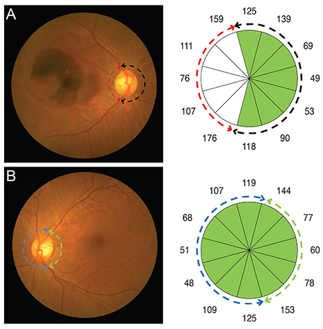
Fig. 1 Fundus photograph and retinal nerve fiber layer thickness analysis of (A) the right affected eye and (B) the left normal fellow eye of a patient with age-related macular degeneration. The red and black arrows represent the pathologic and non-pathologic areas in the affected eye, respectively. The green and blue arrows represent the same area in the unaffected eye, respectively.

Fig. 2 Longitudinal changes in average and quadrant retinal nerve fiber layer (RNFL) thickness in affected eyes. The differences between baseline and postinjection at 6 months (p = 0.012) and 12 months (p = 0.006) were statistically significant in the temporal areas. *p < 0.05, Wilcoxon signed-rank test.
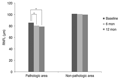
Fig. 3 Longitudinal changes in average retinal nerve fiber layer (RNFL) thickness in the pathologic and non-pathologic areas of affected eyes. The differences between baseline and post-injection at 6 months (p = 0.011) and 12 months (p = 0.005) were statistically significant in the pathologic areas. *p < 0.05, Wilcoxon signed-rank test.
Reference
-
1. Rosenfeld PJ, Brown DM, Heier JS, et al. Ranibizumab for neovascular age-related macular degeneration. N Engl J Med. 2006; 355:1419–1431.2. Gunther JB, Altaweel MM. Bevacizumab (Avastin) for the treatment of ocular disease. Surv Ophthalmol. 2009; 54:372–400.3. el Matri L, Chebil A, Kort F, et al. Intravitreal injection of triamcinolone combined with bevacizumab for choroidal neovascularization associated with large retinal pigment epithelial detachment in age-related macular degeneration. Graefes Arch Clin Exp Ophthalmol. 2010; 248:779–784.4. Jager RD, Aiello LP, Patel SC, Cunningham ET Jr. Risks of intravitreous injection: a comprehensive review. Retina. 2004; 24:676–698.5. Angulo Bocco MC, Glacet-Bernard A, Zourdani A, et al. Intravitreous injection: retrospective study on 2028 injections and their side effects. J Fr Ophtalmol. 2008; 31:693–698.6. Sampat KM, Garg SJ. Complications of intravitreal injections. Curr Opin Ophthalmol. 2010; 21:178–183.7. Day S, Acquah K, Mruthyunjaya P, et al. Ocular complications after anti-vascular endothelial growth factor therapy in Medicare patients with age-related macular degeneration. Am J Ophthalmol. 2011; 152:266–272.8. Hollands H, Wong J, Bruen R, et al. Short-term intraocular pressure changes after intravitreal injection of bevacizumab. Can J Ophthalmol. 2007; 42:807–811.9. Falkenstein IA, Cheng L, Freeman WR. Changes of intraocular pressure after intravitreal injection of bevacizumab (Avastin). Retina. 2007; 27:1044–1047.10. Kim JE, Mantravadi AV, Hur EY, Covert DJ. Short-term intraocular pressure changes immediately after intravitreal injections of anti-vascular endothelial growth factor agents. Am J Ophthalmol. 2008; 146:930–934.e1.11. Sharei V, Hohn F, Kohler T, et al. Course of intraocular pressure after intravitreal injection of 0.05 mL ranibizumab (Lucentis). Eur J Ophthalmol. 2010; 20:174–179.12. Kahook MY, Kimura AE, Wong LJ, at al. Sustained elevation in intraocular pressure associated with intravitreal bevacizumab injections. Ophthalmic Surg Lasers Imaging. 2009; 40:293–295.13. Bakri SJ, McCannel CA, Edwards AO, Moshfeghi DM. Persisent ocular hypertension following intravitreal ranibizumab. Graefes Arch Clin Exp Ophthalmol. 2008; 246:955–958.14. Adelman RA, Zheng Q, Mayer HR. Persistent ocular hypertension following intravitreal bevacizumab and ranibizumab injections. J Ocul Pharmacol Ther. 2010; 26:105–110.15. Jalil A, Fenerty C, Charles S. Intravitreal bevacizumab (Avastin) causing acute glaucoma: an unreported complication. Eye (Lond). 2007; 21:1541.16. Sondell M, Lundborg G, Kanje M. Vascular endothelial growth factor has neurotrophic activity and stimulates axonal outgrowth, enhancing cell survival and Schwann cell proliferation in the peripheral nervous system. J Neurosci. 1999; 19:5731–5740.17. Zachary I. Neuroprotective role of vascular endothelial growth factor: signalling mechanisms, biological function, and therapeutic potential. Neurosignals. 2005; 14:207–221.18. Nishijima K, Ng YS, Zhong L, et al. Vascular endothelial growth factor-A is a survival factor for retinal neurons and a critical neuroprotectant during the adaptive response to ischemic injury. Am J Pathol. 2007; 171:53–67.19. Horsley MB, Mandava N, Maycotte MA, Kahook MY. Retinal nerve fiber layer thickness in patients receiving chronic anti-vascular endothelial growth factor therapy. Am J Ophthalmol. 2010; 150:558–561.e1.20. Seth RK, Salim S, Shields MB, Adelman RA. Assessment of optic nerve cup-to-disk ratio changes in patients receiving multiple intravitreal injections of antivascular endothelial growth factor agents. Retina. 2009; 29:956–959.21. Martinez-de-la-Casa JM, Ruiz-Calvo A, Saenz-Frances F, et al. Retinal nerve fiber layer thickness changes in patients with age-related macular degeneration treated with intravitreal ranibizumab. Invest Ophthalmol Vis Sci. 2012; 53:6214–6218.22. Hwang DJ, Lee EJ, Lee SY, et al. Effect of diabetic macular edema on peripapillary retinal nerve fiber layer thickness profiles. Invest Ophthalmol Vis Sci. 2014; 55:4213–4219.
- Full Text Links
-
- Actions
-
Cited
- CITED
-
- Close
- Share
-
- Similar articles
-
- Ganglion Cell Layer Thickness after Anti-Vascular Endothelial Growth Factor Treatment in Retinal Vein Occlusion
- Anti-Vascular Endothelial Growth Factor Therapy for Choroidal Neovascularization Secondary to Optic Nerve Head Drusen
- Reproducibility of Retinal Nerve Fiber Layer Thickness Evaluation by Nerve Fiber Analyzer
- Treatment of Exudative Age-Related Macular Degeneration
- Intravitreal injection of anti-vascular endothelial growth factor for patients with various retinal diseases
