Obstet Gynecol Sci.
2016 Mar;59(2):91-96. 10.5468/ogs.2016.59.2.91.
Simple mathematical formulae for estimation of median values of fetal biometry at each gestational age
- Affiliations
-
- 1Department of Obstetrics and Gynecology, Seoul National University College of Medicine, Seoul, Korea.
- 2Department of Obstetrics and Gynecology, Seoul National University Bundang Hospital, Seongnam, Korea. hjsobgy@gmail.com
- KMID: 2159002
- DOI: http://doi.org/10.5468/ogs.2016.59.2.91
Abstract
OBJECTIVE
The aim of this study was to propose simple mathematical formulae to estimate median values of fetal biometry including biparietal diameter (BPD), abdominal circumference (AC) and femur length (FL) at each gestational age (GA) easily without looking up the previously established reference values.
METHODS
Simple mathematical formulae to estimate median values of fetal biometric values at each gestational week were inferred. To validate these formulae, three different linear equations were derived from previously reported reference values of median BPD, AC and FL using regression analysis at each gestational week. Finally, calculated data through the inferred formula were compared to retrospectively collected data (observed data).
RESULTS
The equation revealing the relationship between BPD and GA was: median BPD (cm)=GA (wk)/4. Using this simple mathematical formula, the absolute percentage error between observed data and calculated data ranged from 0.12% to 7.50%. The equation between AC and GA was: median AC (cm)=GA (wk)-5. Through this formula, the absolute percentage error was analyzed same as above and it ranged from 0.30% to 4.76%. Lastly the derived formula between FL and GA was: median FL (cm)=GA (wk)/5 and the absolute percentage error ranged from 4.52% to 16.75%.
CONCLUSION
The three simple formulae suggested in our study showed a significantly easy way to estimate the median values of fetal biometry at each gestational week with good reliability.
Keyword
Figure
-
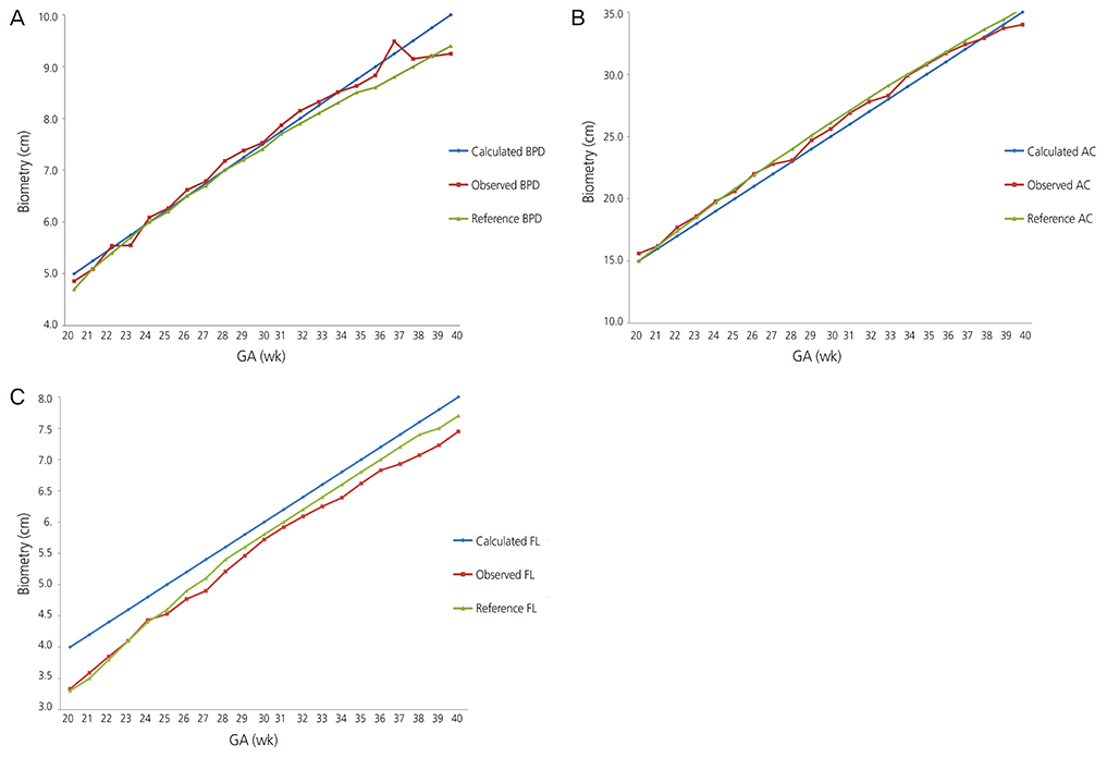
Fig. 1 Schematic comparison between observed, calculated and reference data in graphs. (A), (B), and (C) depict values of calculated, observed and reference values of fetal biparietal diameter (BPD), abdominal circumference (AC) and femur length (FL), respectively. The figures show that the values are quite approximate throughout all gestational weeks (after gestational age [GA] 20 weeks).
Reference
-
1. Fox HE, Hohler CW. Fetal evaluation by real-time imaging. Clin Obstet Gynecol. 1977; 20:339–349.2. Cunningham FG, Williams JW. Williams obstetrics. 24th ed. New York (NY): McGrawHill;2014.3. Hadlock FP, Harrist RB, Martinez-Poyer J. In utero analysis of fetal growth: a sonographic weight standard. Radiology. 1991; 181:129–133.4. Hadlock FP, Deter RL, Harrist RB, Park SK. Computer assisted analysis of fetal age in the third trimester using multiple fetal growth parameters. J Clin Ultrasound. 1983; 11:313–316.5. Hadlock FP, Harrist RB, Sharman RS, Deter RL, Park SK. Estimation of fetal weight with the use of head, body, and femur measurements: a prospective study. Am J Obstet Gynecol. 1985; 151:333–337.6. Jung SI, Lee YH, Moon MH, Song MJ, Min JY, Kim JA, et al. Reference charts and equations of Korean fetal biometry. Prenat Diagn. 2007; 27:545–551.7. Lee JJ. Birth weight for gestational age patterns by sex, plurality, and parity in Korean population. Korean J Pediatr. 2007; 50:732–739.8. Yaghoobian J. Simplified method for estimation of gestational age by biparietal diameter measurement. J Diagn Med Sonogr. 1987; 3:33–35.9. Honarvar M, Allahyari M, Dehbashi S. Assessment of gestational age based on ultrasonic femur length after the first trimester: a simple mathematical correlation between gestational age (GA) and femur length (FL). Int J Gynaecol Obstet. 2000; 70:335–340.10. Rosati P, Guariglia L. Mathematical models to predict long bone lengths by transvaginal scan at 11-16 weeks' gestation. Fetal Diagn Ther. 2003; 18:51–53.11. Callen PW. Ultrasonography in obstetrics and gynecology. 5th ed. Philadelphia (PA): Saunders Elsevier;2008.12. Bertino E, Di Battista E, Bossi A, Pagliano M, Fabris C, Aicardi G, et al. Fetal growth velocity: kinetic, clinical, and biological aspects. Arch Dis Child Fetal Neonatal Ed. 1996; 74:F10–F15.13. Rosati P, Guariglia L, Capelli G. A new mathematical formula for predicting long bone length in early pregnancy. Ultrasound Obstet Gynecol. 2002; 19:184–189.
- Full Text Links
-
- Actions
-
Cited
- CITED
-
- Close
- Share
-
- Similar articles
-
- Estimation of Mean Fetal Biometry by Using Ultrasonography in Normal Pregnancy
- Ultrasound Measurement of Korean Fetal Growth Parameters by Gestational Age in Midtrimester Pregnancy
- Estimation of Fetal Growth by Measurement of Birth Weight for Gestational Age in Newborn
- Clinical Estimational of Gestational Age by Means of Neurologic Examination
- The Change of Fetal Liver Length and Liver Volume by Ultra-sonography according to Gestational Age in Normal Pregnancy
