Spontaneous Rupture of the Second and Third Extensor Digitorum Longus Tendons Caused by Osteophyte of the Tarsometatarsal Joint: A Case Report
- Affiliations
-
- 1Department of Orthopedic Surgery, Gyeongsan Joongang Hospital, Gyeongsan, Korea.
- 2Department of Orthopaedic Surgery, Yeungnam University College of Medicine, Daegu, Korea. chpark77@naver.com
- KMID: 2158430
- DOI: http://doi.org/10.14193/jkfas.2016.20.1.46
Abstract
- Spontaneous rupture of the extensor tendon has been reported in association with predisposing inflammatory conditions including rheumatoid arthritis, diabetes, trauma, tophaceous gout, and steroid injection. The authors experienced a case of spontaneous rupture of the extensor digitorum longus tendons caused by an osteophyte of the tarsometatarsal joint in a patient with rheumatoid arthritis. The authors stress that aggressive treatment including surgery could be considered for prevention of spontaneous tendon rupture in a patient with predisposing conditions despite an asymptomatic spur.
MeSH Terms
Figure
-
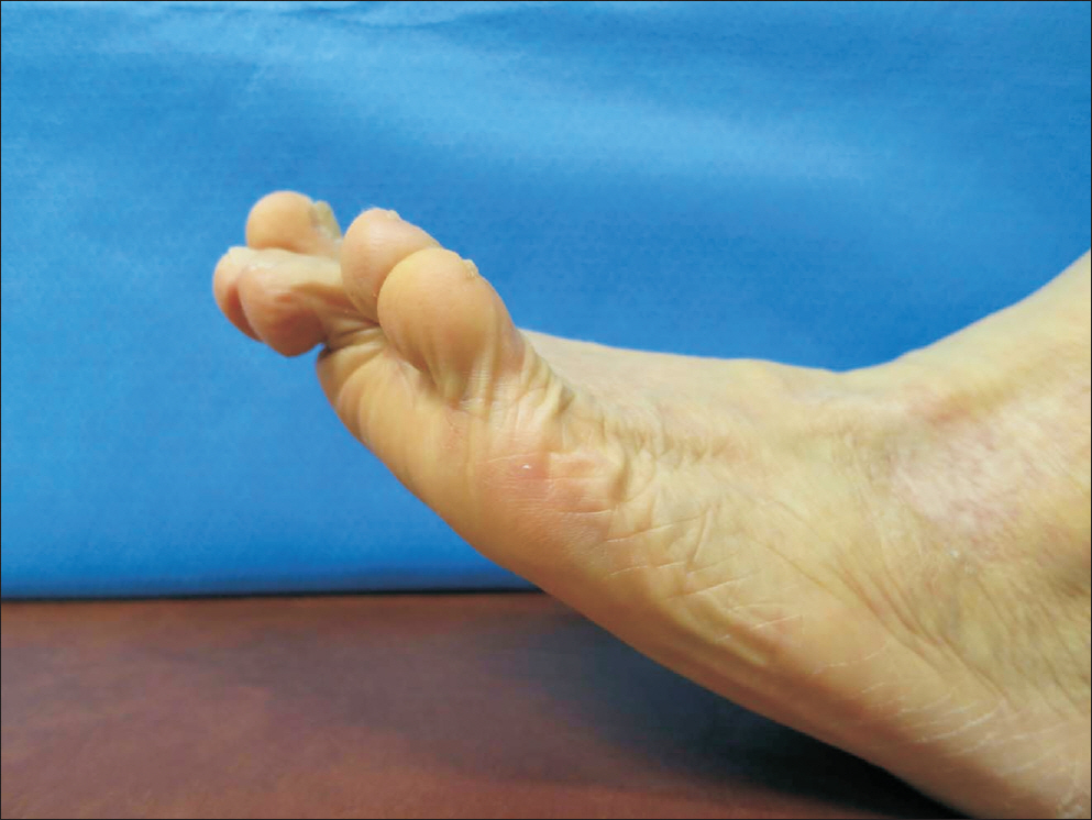
Figure 1. Preoperative photograph shows the patient unable to actively extend the second and third toe.

Figure 2. Preoperative anteroposterior (A) and lateral (B, C) radiographs show flatfoot deformity in both feet, and space narrowing of tarsometatarsal and naviculocuneiform joints with an osteophyte of tarsometatarsal joint in left foot.
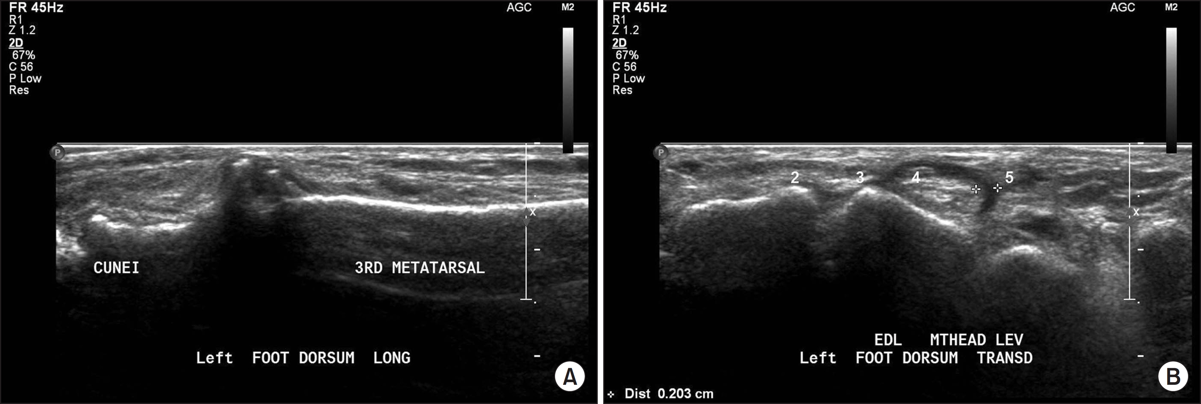
Figure 3. Preoperative ultrasonographs show an osteophyte of tarsometatarsal joint (A) and rupture of second and third extensor digitorum longus tendons (B).

Figure 4. Intraoperative photographs show an osteophyte of tarsometatarsal joint (A), removal of an osteophyte (B), ruptured second and third extensor digitorum longus tendons (C), and repaired tendons using Pulvertaft technique (D).
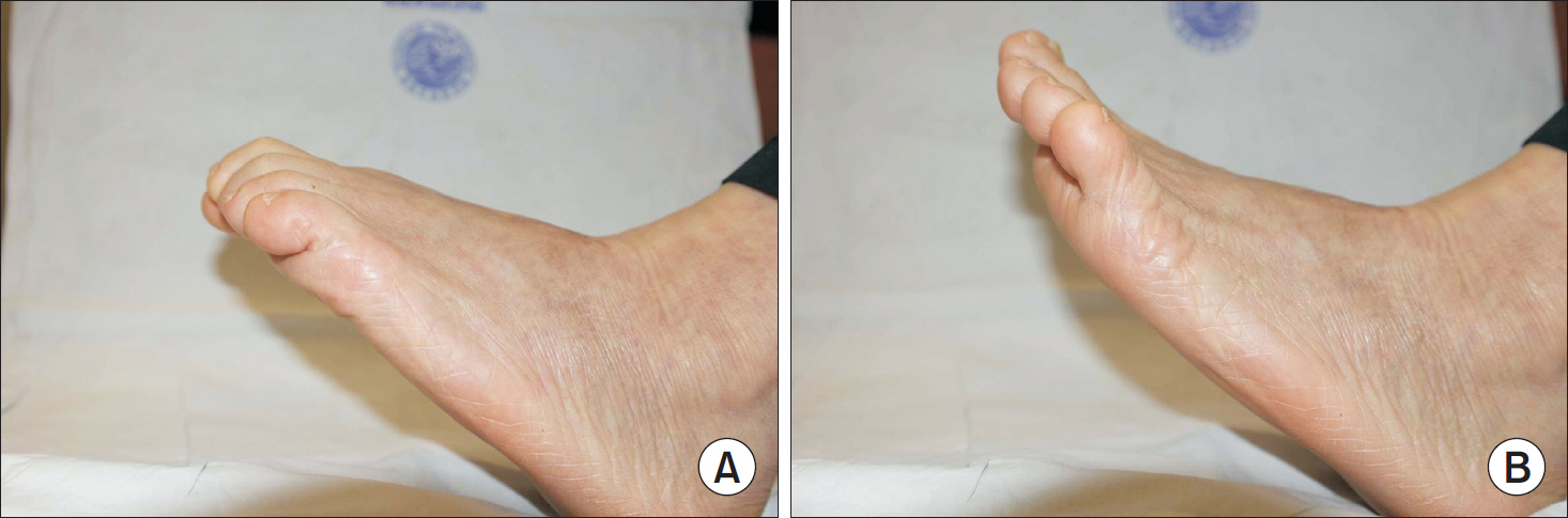
Figure 5. (A, B) Photographs taken at one year after surgery show active range of motion of operated toes.
Reference
-
References
1. Park JW, Kim SK, Park JH, Wang JH, Jeon WJ. Multiple extensor tendon ruptures with advanced Kienboöck's disease. J Hand Surg Am. 2007; 32:233–5.2. Saitoh S, Hata Y, Murakami N, Nakatsuchi Y, Seki H, Takaoka K. Scaphoid nonunion and flexor pollicis longus tendon rupture. J Hand Surg Am. 1999; 24:1211–9.
Article3. Hung JY, Wang SJ, Wu SS. Spontaneous rupture of extensor pollicis longus tendon with tophaceous gout infiltration. Arch Orthop Trauma Surg. 2005; 125:281–4.
Article4. Vaughan-Jackson OJ. Rupture of extensor tendons by attrition at the inferior radioulnar joint; report of two cases. J Bone Joint Surg Br. 1948; 30:528–30.5. Fadel GE, Alipour F. Rupture of the extensor hallucis longus tendon caused by talar neck osteophyte. Foot Ankle Surg. 2008; 14:100–2.
Article6. Pedowitz WJ, Kovatis P. Flatfoot in the Adult. J Am Acad Orthop Surg. 1995; 3:293–302.
Article7. Bouysset M, Tebib J, Noel E, Tavernier T, Miossec P, Vianey JC, et al. Rheumatoid flat foot and deformity of the first ray. J Rheumatol. 2002; 29:903–5.8. Hattori T, Hashimoto J, Tomita T, Kitamura T, Yoshikawa H, Sugamoto K. Radiological study of joint destruction patterns in rheumatoid flatfoot. Clin Rheumatol. 2008; 27:733–7.
Article9. Schindele SF, Herren DB, Simmen BR. Tendon reconstruction for the rheumatoid hand. Hand Clin. 2011; 27:105–13.
Article10. Chung US, Kim JH, Seo WS, Lee KH. Tendon transfer or tendon graft for ruptured finger extensor tendons in rheumatoid hands. J Hand Surg Eur Vol. 2010; 35:279–82.
- Full Text Links
-
- Actions
-
Cited
- CITED
-
- Close
- Share
-
- Similar articles
-
- Spontaneous Rupture of 3rd, 4th, and 5th Extensor Digitorum Tendons in an Amateur Golfer: A Case Report
- Idiopathic Rupture of the Extensor Pollicis Longus Tendon due to Carpometacarpal Joint Arthritis of the Thumb: A Case Report
- Extensor Pollicis Longus Tendon Rupture with Concomitant Rupture of the Extensor Digitorum Communis II Tendon and Extensor Indicis Proprius after Volar Plating for Distal Radius Fracture
- Spontaneous Rupture of the Extensor Pollicis Longus Tendon in a Rhythm Gamer: A Case Report
- Delayed Rupture of the Extensor Pollicis Longus due to Fracture of the distal radius: A Case Report
