A Case of Spitzoid Melanoma with Lymph Node Metastasis in a Child
- Affiliations
-
- 1Department of Dermatology, Korea University College of Medicine, Seoul, Korea. dermaseo@yahoo.co.kr
- KMID: 2157910
- DOI: http://doi.org/10.3346/jkms.2012.27.4.454
Abstract
- The distinction of a spitz nevus from a melanoma can be difficult and in some cases, impossible. A misdiagnosed spitz nevus can metastasize and lead to fatal outcomes, especially in children. A 5-yr-old girl presented with a 1-yr history of a solitary pinkish nodule on her left hand. On physical examination, she had a palpable left axillary lymph node. We performed biopsy and checked 3 sentinel lymph nodes (SLN) on her axillary area. The biopsy specimen showed multiple variably sized and shaped nests with large spindle or polygonal cells and SLN biopsy showed 3 of 3 lymph nodes that were metastasized. Under the diagnosis of spitzoid melanoma, she was treated with excision biopsy and complete left axillary lymph nodes were dissected. She received interferon-alpha2b subcutaneously at a dose of 8 MIU per day, 3 times weekly for 12 months, and shows no recurrence.
Keyword
MeSH Terms
Figure
-
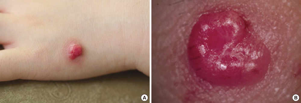
Fig. 1 Gross view of the skin lesion. (A) Slightly tender, solitary pinkish protruding nodule on the lateral side of the dorsum of her left hand. (B) Dermoscopic photo of lesion.
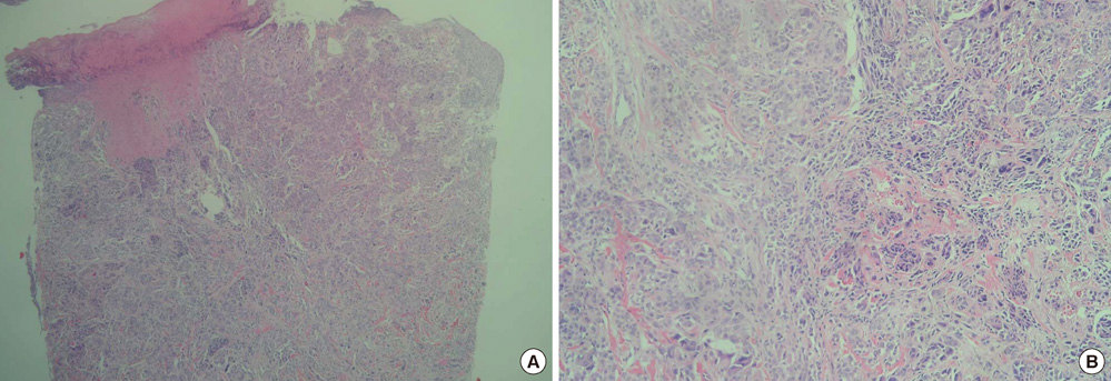
Fig. 2 Histopathology of the skin lesion. (A) Abundant irregular nests of different sizes and shapes and asymmetrical proliferation of enlarged atypical epithelioid and spindle shaped melanocytes in the deep dermis. No maturation of cells from near the surface to the base (H&E stain, × 40). (B) At high power magnification, atypical epithelioid and spindle shaped melanocytes with large, hyperchromatic, pleomorphic nuclei were observed (H&E stain, × 100).
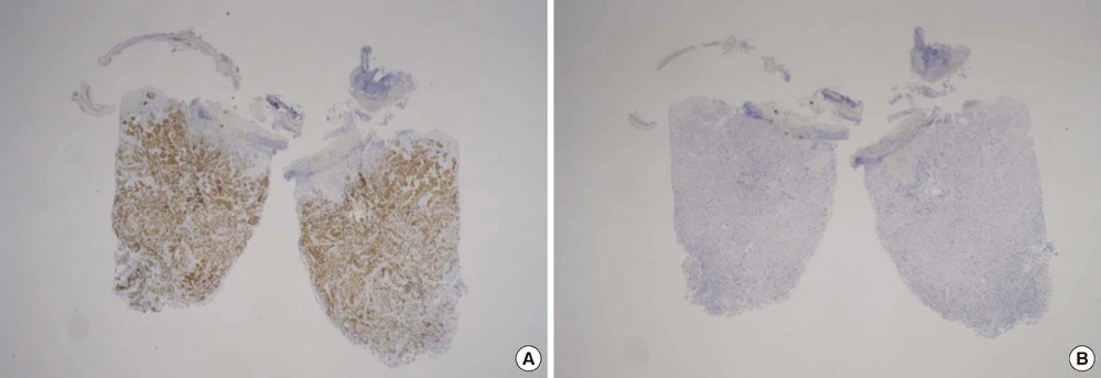
Fig. 3 Immunohistochemistry of the skin lesion. (A) Melan-A is positive at the whole skin level (Melan-A, × 12.5) and (B) weakly positive for HMB-45 (HMB-45, × 12.5).

Fig. 4 Immunohistochemistry of the lymph nodes. (A) The biopsy specimen from the axillary lymph node shows positive results for Melan-A (Melan-A, × 12.5). (B) Atypical melanocytes found within the lymph node parenchyma (Melan-A, × 40).
Cited by 1 articles
-
A Pediatric Case of Spitzoid Melanoma with Subsequent Large Lymph Node Metastasis
Kenji Hayashida, Hiroto Saijo, Shin Morooka, Masaki Fujioka
Ann Dermatol. 2015;27(3):338-339. doi: 10.5021/ad.2015.27.3.338.
Reference
-
1. Kamino H. Spitzoid melanoma. Clin Dermatol. 2009. 27:545–555.2. Elder DE, Elenitsas R, Murphy GF, Xu X. Elder D, Elenitsas R, Johnson BL, Murphy GF, Xu X, editors. Benign pigmented lesions and malignant melanoma. Lever's histopathology of the skin. 2009. 10th ed. Philadelphia: Lippincott Williams & Wilkins;718–724.3. Paradela S, Fonseca E, Pita S, Kantrow SM, Goncharuk VN, Diwan H, Prieto VG. Spitzoid melanoma in children: clinicopathological study and application of immunohistochemistry as an adjunct diagnostic tool. J Cutan Pathol. 2009. 36:740–752.4. Barnhill RL, Gupta K. Unusual variants of malignant melanoma. Clin Dermatol. 2009. 27:564–587.5. Pol-Rodriquez M, Lee S, Silvers DN, Celebi JT. Influence of age on survival in childhood spitzoid melanomas. Cancer. 2007. 109:1579–1583.6. Lee DA, Cohen JA, Twaddell WS, Palacios G, Gill M, Levit E, Halperin AJ, Mones J, Busam KJ, Silvers DN, Celebi JT. Are all melanomas the same? Spitzoid melanoma is a distinct subtype of melanoma. Cancer. 2006. 106:907–913.7. Berk DR, LaBuz E, Dadras SS, Johnson DL, Swetter SM. Melanoma and melanocytic tumors of uncertain malignant potential in children, adolescents and young adults: the Stanford experience 1995-2008. Pediatr Dermatol. 2010. 27:244–254.8. Bastian BC, Wesselman U, Pinkel D, Leboit PE. Molecular cytogenetic analysis of Spitz nevi shows clear differences to melanoma. J Invest Dermatol. 1999. 113:1065–1069.9. Bastian BC, LeBoit PE, Pinkel D. Mutations and copy number increases of HRAS in Spitz nevi with distinctive histopathological features. Am J Pathol. 2000. 157:967–972.10. Bergman R, Malkin L, Sabo E, Kerner H. MIB-1 monoclonal antibody to determine proliferative activity of Ki-67 antigen as an adjunct to the histopathologic differential diagnosis of Spitz nevi. J Am Acad Dermatol. 2001. 44:500–504.11. Vollmer RT. Use of Bayes rule and MIB-1 proliferation index to discriminate Spitz nevus from malignant melanoma. Am J Clin Pathol. 2004. 122:499–505.12. Murali R, Sharma RN, Thompson JF, Stretch JR, Lee CS, McCarthy SW, Scolyer RA. Sentinel lymph node biopsy in histologically ambiguous melanocytic tumors with spitzoid features (so-called atypical spitzoid tumors). Ann Surg Oncol. 2008. 15:302–309.13. Mooi WJ, Krausz T. Spitz nevus versus spitzoid melanoma: diagnostic difficulties, conceptual controversies. Adv Anat Pathol. 2006. 13:147–156.14. Busam KJ, Murali R, Pulitzer M, McCarthy SW, Thompson JF, Shaw HM, Brady MS, Coit DG, Dusza S, Wilmott J, Kayton M, Laquaglia M, Scolyer RA. Atypical spitzoid melanocytic tumors with positive sentinel lymph nodes in children and teenagers, and comparison with histologically unambiguous and lethal melanomas. Am J Surg Pathol. 2009. 33:1386–1395.15. Busam KJ, Pulitzer M. Sentinel lymph node biopsy for patients with diagnostically controversial Spitzoid melanocytic tumors? Adv Anat Pathol. 2008. 15:253–262.
- Full Text Links
-
- Actions
-
Cited
- CITED
-
- Close
- Share
-
- Similar articles
-
- A Pediatric Case of Spitzoid Melanoma with Subsequent Large Lymph Node Metastasis
- Treatment of Malignant Melanoma Using Sentinel Lymph Node Dissection
- A Case of Spitzoid Melanoma
- A Case of Thin Acral Lentiginous Melanoma with Lymph Node Metastasis and Regression
- Popliteal Lymph Node Dissection in Lower Extremity Malignant Melanoma
