J Korean Med Sci.
2006 Aug;21(4):778-780. 10.3346/jkms.2006.21.4.778.
Intradural Disc Herniation at L5-S1 Mimicking an Intradural Extramedullary Spinal Tumor: A Case Report
- Affiliations
-
- 1Department of Orthopedic Surgery, Pusan National University School of Medicine, Busan, Korea. kuentak@pusan.ac.kr
- KMID: 2157840
- DOI: http://doi.org/10.3346/jkms.2006.21.4.778
Abstract
- Intradural lumbar disc herniation is a rare pathological entity. The pathogenesis of intradural lumbar disc herniation is not known clearly. Intradural disc herniations usually occurred at the L4-L5 levels but have also been reported at other levels. However, intradural disc herniation at L5-S1 is quite rare. There are approximately nine reports in the English literature of intraradicular disc herniation at L5-S1. We described a 61-yr-old man with suspected intradural mass at the level of L5-S1 space. The patient presented with pain in the lower back and both lower legs for 4 months and a sudden exacerbation of the symptoms for 3 days. Gadolinium-enhanced magnetic resonance imaging (MRI) demonstrated a large disc herniation at the L5-S1 level with an intradural component. L5 and S1 laminectomy was performed, and dura was swollen and immobile. Subsequent durotomy was performed and an intradural disc fragment was removed. The patient had full recovery in 3 months. Intradural lumbar disc herniation must be considered in the differential diagnosis of mass lesions in the spinal canal. Contrast-enhanced MRI scans are useful to differentiate a herniated disc from a disc space infection or tumor.
Keyword
MeSH Terms
Figure
-
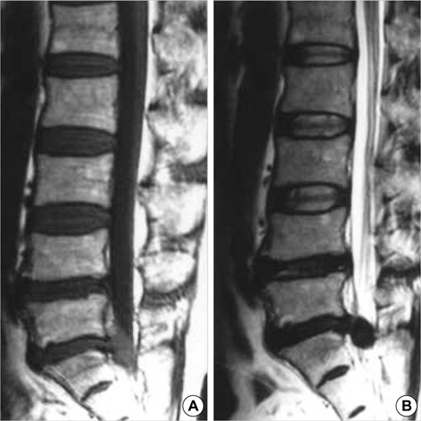
Fig. 1 T1-(A) and T2-weighted (B) sagittal magnetic resonance images demonstrating a mass-like lesion.
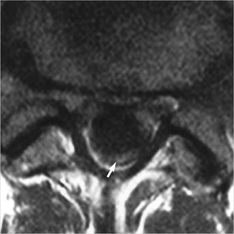
Fig. 2 Contrast-enhanced axial image showing peripheral enhancement of the lesion (arrow).

Fig. 3 Intraoperative photograph (A) outlining the peripheral displacement of the adherent cauda equine nerve roots (arrows) by the large intradural disc fragment. Intraoperative photograph (B) showing an anterior dural defect.
Reference
-
1. Epstein NE, Syrquin MS, Epstein JA, Decker RE. Intradural disc herniations in the cervical, thoracic, and lumbar spine: report of three cases and review of the literature. J Spinal Disord. 1990. 3:396–403.2. D'Andrea G, Trillò G, Roperto R, Celli P, Orlando ER, Ferrante L. Intradural lumbar disc herniations: the role of MRI in preoperative diagnosis and review of the literature. Neurosurg Rev. 2004. 27:75–80.3. Mut M, Berker M, Palaoglu S. Intraradicular disc herniations in the lumbar spine and a new classification of intradural disc herniations. Spinal Cord. 2001. 39:545–548.
Article4. Aydin MV, Ozel S, Sen O, Erdogan B, Yildirim T. Intradural disc mimicking a spinal tumor lesion. Spinal Cord. 2004. 42:52–54.
Article5. Suzer T, Tahta K, Coskun E. Intraradicular lumbar disc herniation: Case report and review of the literature. Neurosurgery. 1997. 41:956–959.6. Tsuji H, Maruta K, Maeda A. Postoperative intraradicular intervertebral disc herniation. Spine. 1991. 61:998–1000.
Article7. Blikra G. Intradural herniated lumbar disc. J Neurosurg. 1969. 31:676–679.
Article8. Spencer DL, Irwin GS, Miller JA. Anatomy and significance of fixation of the lumbosacral nerve roots in sciatica. Spine. 1983. 8:672–679.
Article9. Lidov M, Stollman A, Casden A, Som P, Bederson J. MRI of lumbar intradural disc herniation. Clin Imaging. 1994. 18:173–178.
Article
- Full Text Links
-
- Actions
-
Cited
- CITED
-
- Close
- Share
-
- Similar articles
-
- Lumbar Intradural Disc Herniation Mimicking a Chondroma
- Huge Intradural Lumbar Disc Herniation Mimicking an Intradural Spinal Tumor: A Case Report
- Multiple Intradural Disc Herniations Masquerading as Intradural Extramedullary Tumors: A Case Report and Review of the Literature
- Intradural Migration of a Sequestrated Lumbar Disc Fragment Masquerading as a Spinal Intradural Tumor
- A Case Report of Intradural Ruptured Lumbar Disc
