Usefulness of Dermatoscopy for the Preoperative Assessment of the Histopathologic Aggressiveness of Basal Cell Carcinoma
- Affiliations
-
- 1Department of Dermatology, Pusan National University School of Medicine, Busan, Korea. drkmp@hanmail.net
- 2Biomedical Research Institute, Pusan National University Hospital, Busan, Korea.
- KMID: 2157443
- DOI: http://doi.org/10.5021/ad.2015.27.6.682
Abstract
- BACKGROUND
Limited information is available regarding dermatoscopic differences between non-aggressive and aggressive types of basal cell carcinoma (BCC).
OBJECTIVE
To investigate dermatoscopic differences between non-aggressive and aggressive types.
METHODS
We evaluated 145 histopathologically confirmed BCCs from 141 patients. Histopathologic types and aggressiveness from 4 mm punch biopsy and their dermatoscopic findings were evaluated. We assessed the statistical significance of dermatoscopic differences between non-aggressive and aggressive types. To objectively predict aggressiveness, we created a "dermatoscopic index of BCC aggressiveness" in which 1 point was added and subtracted for each dermatoscopic finding significantly higher in aggressive and non-aggressive types, respectively.
RESULTS
Large blue-gray ovoid nests were found more frequently in non-aggressive type than aggressive one (85/105 [80.9%] vs. 21/40 [52.5%], p=0.001). Compared to non-aggressive type, aggressive type had more multiple blue-gray globules (29/40 [72.5%] vs. 57/105 [54.3%], p=0.046), arborizing telangiectasia (29/40 [72.5%] vs. 48/105 [45.7%], p=0.004), and concentric structure (11/40 [27.5%] vs. 12/105 [11.4%], p=0.018). Regarding dermatoscopic index, cases of aggressive type with a score of 1 were most common (n=18, 45.0%), followed by a score of 2 (n=14, 35.0%). Limited number of aggressive type of BCCs and the effect of width on the determination of histopathologic aggressiveness.
CONCLUSION
Aggressive type BCCs more often exhibited multiple blue-gray globules, arborizing telangiectasia, and concentric structure, while the non-aggressive type exhibited large blue-gray ovoid nests more frequently. Score exceeding 2 on the dermoscopic index can be screening criteria for aggressiveness. These dermatoscopic features and dermoscopic index could be useful for assessing aggressiveness of BCCs before surgery.
Figure
-
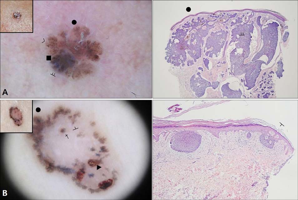
Fig. 1 Non-aggressive types of basal cell carcinoma. (A) Nodular type, (B) superficial type. Left column: dermatoscopic findings (inset, clinical photo). Right column: histologic findings (H&E, ×40). ᚇ: large blue-gray ovoid nests, ■: multiple blue-gray globules, ●: maple leaf-like areas, ᚆ: spoke-wheel areas, ▲: ulceration, †: concentric structure, >: brown-black dots.
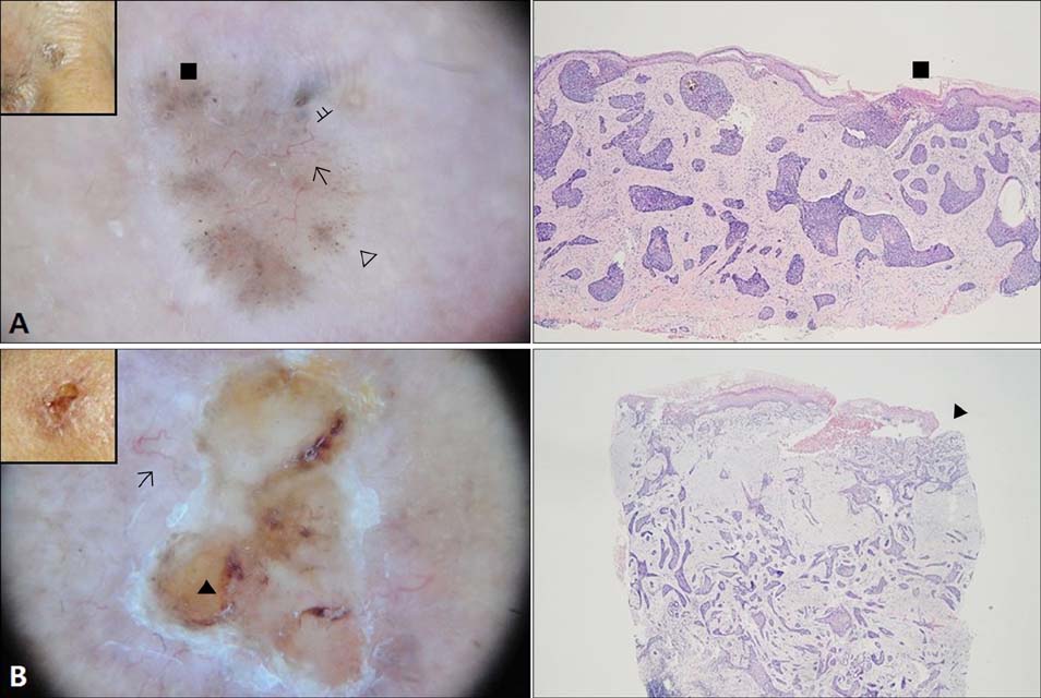
Fig. 2 Aggressive types of basal cell carcinoma. (A) Micronodular type, (B) infiltrative type. Left column: dermatoscopic findings (inset, clinical photo). Right column: histologic findings (H&E, ×40). ᚇ: large blue-gray ovoid nests, ■: multiple blue-gray globules, ➔: arborizing telangiectasia, ▲: ulceration, △: multiple blue-gray dots.
Cited by 1 articles
-
Increased Immunoreactivity of LGR4 in Histologically Aggressive Basal Cell Carcinoma
Shin-Taek Oh, Junguee Lee, Keum-Jin Yang, Jung-Min Bae, Hyun-Jeong Park, Jin-Woo Kim, Young-Min Park
Ann Dermatol. 2018;30(5):630-633. doi: 10.5021/ad.2018.30.5.630.
Reference
-
1. Wolberink EA, Pasch MC, Zeiler M, van Erp PE, Gerritsen MJ. High discordance between punch biopsy and excision in establishing basal cell carcinoma subtype: analysis of 500 cases. J Eur Acad Dermatol Venereol. 2013; 27:985–989.
Article2. Kim DH, Kwon IH, Cho KH. A statistical study of cutaneous malignant tumors (2001~2005). Korean J Dermatol. 2008; 46:1581–1587.3. Lang PG Jr, Maize JC. Histologic evolution of recurrent basal cell carcinoma and treatment implications. J Am Acad Dermatol. 1986; 14:186–196.
Article4. Sexton M, Jones DB, Maloney ME. Histologic pattern analysis of basal cell carcinoma. Study of a series of 1039 consecutive neoplasms. J Am Acad Dermatol. 1990; 23:1118–1126.5. Saldanha G, Fletcher A, Slater DN. Basal cell carcinoma: a dermatopathological and molecular biological update. Br J Dermatol. 2003; 148:195–202.
Article6. Berk DR, Ball Arefiev KL, Gladstone HB. Adamantinoid basal cell carcinoma: a predictor of more-aggressive clinical behavior. Dermatol Surg. 2012; 38:1346–1350.
Article7. Husein-Elahmed H, Aneiros-Fernandez J, Gutierrez-Salmerón MT, Botella-Lopez M, Aneiros-Cachaza J, Naranjo-Sintes R. Alcohol intake and risk of aggressive histological basal cell carcinoma: a case-control study. Eur J Dermatol. 2012; 22:525–530.
Article8. Cohen PR, Schulze KE, Nelson BR. Basal cell carcinoma with mixed histology: a possible pathogenesis for recurrent skin cancer. Dermatol Surg. 2006; 32:542–551.
Article9. Menzies SW, Westerhoff K, Rabinovitz H, Kopf AW, McCarthy WH, Katz B. Surface microscopy of pigmented basal cell carcinoma. Arch Dermatol. 2000; 136:1012–1016.
Article10. Altamura D, Menzies SW, Argenziano G, Zalaudek I, Soyer HP, Sera F, et al. Dermatoscopy of basal cell carcinoma: morphologic variability of global and local features and accuracy of diagnosis. J Am Acad Dermatol. 2010; 62:67–75.
Article11. Peris K, Altobelli E, Ferrari A, Fargnoli MC, Piccolo D, Esposito M, et al. Interobserver agreement on dermoscopic features of pigmented basal cell carcinoma. Dermatol Surg. 2002; 28:643–645.
Article12. Tabanlıoğlu Onan D, Sahin S, Gököz O, Erkin G, CakXMLLink_XYZr B, Elçin G, et al. Correlation between the dermatoscopic and histopathological features of pigmented basal cell carcinoma. J Eur Acad Dermatol Venereol. 2010; 24:1317–1325.
Article13. Giacomel J, Zalaudek I. Dermoscopy of superficial basal cell carcinoma. Dermatol Surg. 2005; 31:1710–1713.
Article14. Micantonio T, Gulia A, Altobelli E, Di Cesare A, Fidanza R, Riitano A, et al. Vascular patterns in basal cell carcinoma. J Eur Acad Dermatol Venereol. 2011; 25:358–361.
Article15. Argenziano G, Soyer HP, Giorgio VD, Piccolo D, Carli P, Delfino M, et al. Interactive atlas of dermoscopy. Milan, Italy: EDRA Medical Publishing & New Media;2002.16. Braun RP, Rabinovitz HS, Oliviero M, Kopf AW, Saurat JH. Dermoscopy of pigmented skin lesions. J Am Acad Dermatol. 2005; 52:109–121.
Article17. Welsch MJ, Troiani BM, Hale L, DelTondo J, Helm KF, Clarke LE. Basal cell carcinoma characteristics as predictors of depth of invasion. J Am Acad Dermatol. 2012; 67:47–53.
Article18. Haws AL, Rojano R, Tahan SR, Phung TL. Accuracy of biopsy sampling for subtyping basal cell carcinoma. J Am Acad Dermatol. 2012; 66:106–111.
Article19. Russell EB, Carrington PR, Smoller BR. Basal cell carcinoma: a comparison of shave biopsy versus punch biopsy techniques in subtype diagnosis. J Am Acad Dermatol. 1999; 41:69–71.
Article20. Newell B, Bedlow AJ, Cliff S, Drysdale SB, Stanton AW, Mortimer PS. Comparison of the microvasculature of basal cell carcinoma and actinic keratosis using intravital microscopy and immunohistochemistry. Br J Dermatol. 2003; 149:105–110.
Article
- Full Text Links
-
- Actions
-
Cited
- CITED
-
- Close
- Share
-
- Similar articles
-
- Dermoscopic and Histopathologic Analysis of the Correlation between the Pigmentation of Basal Cell Carcinoma and Tumor Aggressiveness
- Immunohistochemical Study for Syndecan-1 and Beta-catenin Expression in Basal Cell Carcinoma
- A Case of Adenoid Type Basal Cell Carcinoma on Philtrum
- A case of adenoid basal cell carcinoma in uterine cervix
- A Case of Multiple Basal Cell Carcinomas in Nevoid Basal Cell Carcinoma Syndrome
