Ann Dermatol.
2012 Feb;24(1):94-98. 10.5021/ad.2012.24.1.94.
Effects of Murine Dermal Cells on the Regulation of Hair Growth Is Dependent on the Cell Number and Post-Natal Age of Newborn Mice
- Affiliations
-
- 1Department of Dermatology, Seoul National University College of Medicine, Seoul, Seoul National University Bundang Hospital, Seongnam, Korea. chhuh@snu.ac.kr
- KMID: 2156866
- DOI: http://doi.org/10.5021/ad.2012.24.1.94
Abstract
- Dermal cells from neonatal mice can initiate the formation of hair follicles (HFs) when combined with adult mouse epidermal cells and transplanted subcutaneously into athymic mice. In the present study, the effects of dermal cells on HF formation were tested in terms of total cell number and the time course of cell harvest. Results demonstrated that the number of dermal cells is critical to the formation of HF. Furthermore, hair forming ability is rapidly decreasing as the neonatal mice age. To examine potential differences in gene expression, cDNA array was performed. Results demonstrate that numerous molecules which are directly involved in receptor and signaling correlated with decreased hair inductivity in early time points after delivery. It is reported that bone morphogenic protein (BMP)-6 and Wnt3a treatment increased hair inductivity of dermal papilla cells. But in our study, no changes were observed in the expression levels of BMP-6 and Wnt3a. However, several Wnt related genes demonstrate increased or decreased expression levels. Thus, our results suggest that co-ordinated regulation of these molecules will be important in hair neogenesis within our model system.
Keyword
MeSH Terms
Figure
-
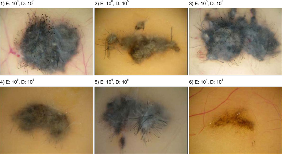
Fig. 1 Formation of nodules at inoculation sites according to different combination of dermal (105~106 cells) and epidermal cells (104~106 cells).

Fig. 2 Histological analysis of nodules according to different combination of dermal and epidermal cells (H&E, ×100).
Cited by 1 articles
-
Poor Capability of 3D-Cultured Adipose-Derived Stem Cells to Induce Hair Follicles in Contrast to 3D-Cultured Dermal Papilla Cells
Chang Hoon Seo, Mi Hee Kwack, Soo-Hong Lee, Moon Kyu Kim, Jung Chul Kim, Young Kwan Sung
Ann Dermatol. 2016;28(5):662-665. doi: 10.5021/ad.2016.28.5.662.
Reference
-
1. Cotsarelis G. Epithelial stem cells: a folliculocentric view. J Invest Dermatol. 2006. 126:1459–1468.
Article2. Yang CC, Cotsarelis G. Review of hair follicle dermal cells. J Dermatol Sci. 2010. 57:2–11.
Article3. Chuong CM, Cotsarelis G, Stenn K. Defining hair follicles in the age of stem cell bioengineering. J Invest Dermatol. 2007. 127:2098–2100.
Article4. Osada A, Iwabuchi T, Kishimoto J, Hamazaki TS, Okochi H. Long-term culture of mouse vibrissal dermal papilla cells and de novo hair follicle induction. Tissue Eng. 2007. 13:975–982.
Article5. Rendl M, Polak L, Fuchs E. BMP signaling in dermal papilla cells is required for their hair follicle-inductive properties. Genes Dev. 2008. 22:543–557.
Article6. Kishimoto J, Burgeson RE, Morgan BA. Wnt signaling maintains the hair-inducing activity of the dermal papilla. Genes Dev. 2000. 14:1181–1185.
Article7. Millar SE. Molecular mechanisms regulating hair follicle development. J Invest Dermatol. 2002. 118:216–225.
Article8. Dai J, Hall CL, Escara-Wilke J, Mizokami A, Keller JM, Keller ET. Prostate cancer induces bone metastasis through Wntinduced bone morphogenetic protein-dependent and independent mechanisms. Cancer Res. 2008. 68:5785–5794.
Article9. Xing Y, Xu W, Yang K, Lian X, Yang T. Immunolocalization of Wnt5a during the hair cycle and its role in hair shaft growth in mice. Acta Histochem. 2011. 113:608–612.
Article10. Katoh M. WNT signaling in stem cell biology and regenerative medicine. Curr Drug Targets. 2008. 9:565–570.
Article
- Full Text Links
-
- Actions
-
Cited
- CITED
-
- Close
- Share
-
- Similar articles
-
- Effects of Alopecia Areata Serum on Proliferation of Cultured Dermal Papilla Cells
- Effect of Chrysanthemum zawadskii Extract on Dermal Papilla Cell Proliferation and Hair Growth
- Effects of Vaseular Endothelial Growth Factors on Hair Growth in Vitro
- Hair Growth Promoting Potential of Phospholipids Purified from Porcine Lung Tissues
- Dermal mast cell responses in Paragonimus westermani-infected mice
