Utility of Dermoscopy before and after Laser Irradiation in Port Wine Stains
- Affiliations
-
- 1Department of Plastic and Reconstructive Surgery, Osaka City University Graduate School of Medicine, Osaka, Japan. m3552667@msic.med.osaka-cu.ac.jp
- KMID: 2156848
- DOI: http://doi.org/10.5021/ad.2012.24.1.7
Abstract
- BACKGROUND
Port wine stains (PWSs) are commonly treated with pulsed dye laser (PDL) as a standard therapy. However, it is not easy to predict the minimal effective dose in the first treatment session.
OBJECTIVE
The aim of this study was to assess whether dermoscopic findings before and after laser irradiation corresponded with the clinical improvement of PWS in patients undergoing PDL therapy.
METHODS
Seven untreated PWSs in 6 patients (a male and 5 females), who presented to our hospital between May 2008 to January 2010, were assessed in this study. The mean age was 36.3 years, ranging from 14 to 57 years. A PDL with a wavelength of 585 nm and a spot size of 7 mm was used. Before and after test irradiation, patients underwent dermoscopy and clinical photography, and we assessed whether the dermoscopic findings corresponded with clinical improvement after 3 months.
RESULTS
There were no obvious differences observed in the clinical photographs between each test level immediately after irradiation. However, dermoscopic photographs showed differences as the irradiated energy increased. These changes corresponded to the clinical improvement after 3 months.
CONCLUSION
Our study indicates that the minimal effective fluence can be predicted by observing dermoscopic change immediately after irradiation. We think that examining the dermoscopic findings immediately after irradiation allows the laser surgeon to predict the minimal effective fluence and this prevents adverse effects of the skin.
Keyword
Figure
-
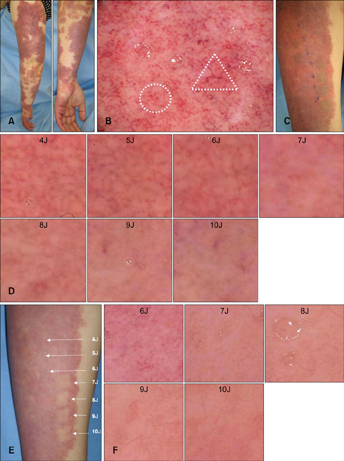
Fig. 1 (A) A 43-year-old woman with a Port wine stain on the left arm before treatment. (B) Dermoscopic photograph before treatment shows (○) dotted and globular vessels (superficial), as well as (▵) linear vessels (deep). (C) Immediately after laser irradiation, there are some differences, such as indistinct margins in 4~5 J/cm2 and increased whiteness in 9~10 J/cm2, but it is difficult to know which is the most effective fluence by the naked eye. (D) Dermoscopic photographs immediately after irradiation (×10). At an energy level of 7 J/cm2, vessel walls are difficult to detect. (E) Three months after irradiation. at an energy level of 7 J/cm2, treatment was effective. (F) Dermoscpic photographs at 3 months after irradiation. at an energy level of 7 J/cm2, vessel walls have disappeared.
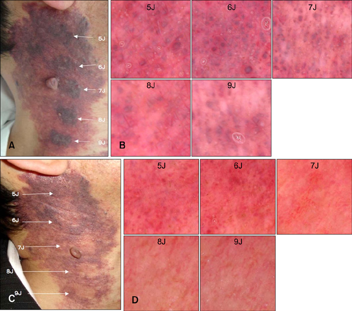
Fig. 2 (A) A 52-year-old man with a previously untreated Port wine stain on the right neck immediately after irradiation. Except for purple change, no obvious difference was seen between each energy level. (B) Dermoscopic photographs obtained immediately after irradiation (×10). At an energy level of 7 J/cm2, whitish areas appear. (C) Three months after irradiation. At an energy level of 7 J/cm2, we evaluated the treatment as effective. (D) Dermoscpic photographs 3 months after irradiation. At an energy level of 7 J/cm2, vessel walls are difficult to detect.
Reference
-
1. Lam AY, Wong DS, Lam LK, Ho WS, Chan HH. A retrospective study on the efficacy and complications of Q-switched alexandrite laser in the treatment of acquired bilateral nevus of Ota-like macules. Dermatol Surg. 2001. 27:937–941.
Article2. Eubanks LE, McBurney EI. Videomicroscopy of port-wine stains: correlation of location and depth of lesion. J Am Acad Dermatol. 2001. 44:948–951.
Article3. Fiskerstrand EJ, Svaasand LO, Kopstad G, Dalaker M, Norvang LT, Volden G. Laser treatment of port wine stains: therapeutic outcome in relation to morphological parameters. Br J Dermatol. 1996. 134:1039–1043.
Article4. Onizuka K, Tsuneda K, Shibata Y, Ito M, Sekine I. Efficacy of flashlamp-pumped pulsed dye laser therapy for port wine stains: clinical assessment and histopathological characteristics. Br J Plast Surg. 1995. 48:271–279.
Article5. Seidenari S, Pellacani G, Pepe P. Digital videomicroscopy improves diagnostic accuracy for melanoma. J Am Acad Dermatol. 1998. 39:175–181.
Article6. Motley RJ, Lanigan SW, Katugampola GA. Videomicroscopy predicts outcome in treatment of port-wine stains. Arch Dermatol. 1997. 133:921–922.
Article7. Procaccini EM, Argenziano G, Staibano S, Ferrara G, Monfrecola G. Epiluminescence microscopy for port-wine stains: pretreatment evaluation. Dermatology. 2001. 203:329–332.
Article8. Suthamjariya K, Farinelli WA, Koh W, Anderson RR. Mechanisms of microvascular response to laser pulses. J Invest Dermatol. 2004. 122:518–525.
Article
- Full Text Links
-
- Actions
-
Cited
- CITED
-
- Close
- Share
-
- Similar articles
-
- A Case of Granuloma Pyogenicum within a Port-wine Stain
- The combined application of bleomycin with triamcinolone acetonide in port wine stains: inhibiting proliferation and recurrence of port wine stains
- Five Cases of Acquired Port-Wine Stains
- Treatment of Port Wine Stain with Nodular Change
- Clinical Observations on Port - Wine Stains
