Ann Dermatol.
2011 Oct;23(Suppl 2):S239-S242. 10.5021/ad.2011.23.S2.S239.
Deep Penetrating Benign Fibrous Histiocytoma of the Foot Associated with Throbbing Pain
- Affiliations
-
- 1Department of Dermatology, Dankook University Hospital, Cheonan, Korea. 4exodus@hanmail.net
- 2Department of Dermatology, Ajou University Hospital, Suwon, Korea.
- 3Department of Dermatology, Eulji University Hospital, Daejeon, Korea.
- KMID: 2156799
- DOI: http://doi.org/10.5021/ad.2011.23.S2.S239
Abstract
- Compared to cutaneous benign fibrous histiocytoma (BFH), deep-seated BFH is very rare and poorly recognized. Both cutaneous and deep-seated BFH are usually asymptomatic. We herein report a 25 year-old woman who presented with a painful mass in her foot that was poorly controlled by analgesics and associated with walking difficulty. After preoperative ultrasonographic evaluation, the mass was completely excised and histologic exam showed spindle cells loosely arranged in storiform architecture, with CD34-, desmin-, S-100-, focal CD68+, vimentin+, smooth muscle actin+, and factor XIIIa+. The patient was diagnosed with deep-seated BFH based on the histologic, radiologic and intraoperative findings.
Keyword
Figure
-
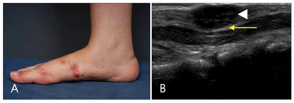
Fig. 1 (A) A painful erythematous nodule was found on the arch of the foot. (B) Well-circumscribed mass from subcutaneous tissue attached to the fascia and compressing the architecture under the fascia (mass: white arrow head, fascia: yellow arrow).
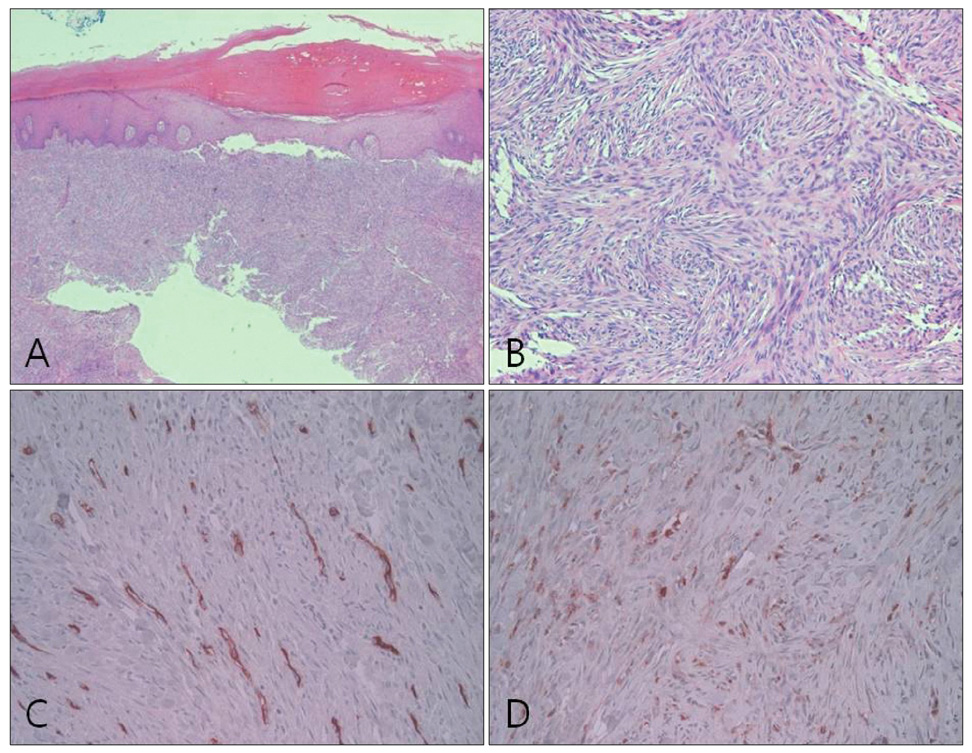
Fig. 2 Loosely arranged spindle cells in storiform architecture, expressing CD34- and CD68+ focally (A: H&E, ×40, B: H&E, ×200, C: CD34, ×200, D: CD68, ×200).
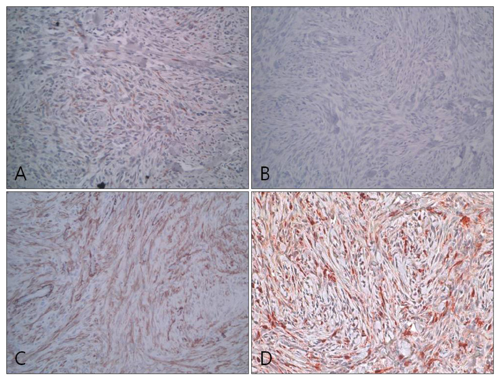
Fig. 3 Tumor cells showed positive reactivity against vimentin, SMA, factor XIIIa, but not against desmin (A: vimentin, ×200, B: desmin, ×200, C: SMA, ×200, D: factor XIIIa, ×200).
Reference
-
1. Skoulakis CE, Papadakis CE, Datseris GE, Drivas EI, Kyrmizakis DE, Bizakis JG. Subcutaneous benign fibrous histiocytoma of the cheek. Case report and review of the literature. Acta Otorhinolaryngol Ital. 2007. 27:90–93.2. Gleason BC, Fletcher CD. Deep "benign" fibrous histiocytoma: clinicopathologic analysis of 69 cases of a rare tumor indicating occasional metastatic potential. Am J Surg Pathol. 2008. 32:354–362.
Article3. Hannachi Sassi S, Trabelsi M, Abid L, Mrad K, Abbess I, Dhouib R, et al. Deep benign fibrous histiocytoma: a case report. Rev Chir Orthop Reparatrice Appar Mot. 2006. 92:809–812.4. Mentzel T, Kutzner H, Rütten A, Hügel H. Benign fibrous histiocytoma (dermatofibroma) of the face: clinicopathologic and immunohistochemical study of 34 cases associated with an aggressive clinical course. Am J Dermatopathol. 2001. 23:419–426.
Article5. Calonje E, Mentzel T, Fletcher CD. Cellular benign fibrous histiocytoma. Clinicopathologic analysis of 74 cases of a distinctive variant of cutaneous fibrous histiocytoma with frequent recurrence. Am J Surg Pathol. 1994. 18:668–676.6. Kaddu S, McMenamin ME, Fletcher CD. Atypical fibrous histiocytoma of the skin: clinicopathologic analysis of 59 cases with evidence of infrequent metastasis. Am J Surg Pathol. 2002. 26:35–46.7. Fletcher CD. Benign fibrous histiocytoma of subcutaneous and deep soft tissue: a clinicopathologic analysis of 21 cases. Am J Surg Pathol. 1990. 14:801–809.
- Full Text Links
-
- Actions
-
Cited
- CITED
-
- Close
- Share
-
- Similar articles
-
- Comments to "Deep Benign Fibrous Histiocytoma Showing Multiple Metastases"
- Deep Benign Fibrous Histiocytoma Showing Multiple Metastases
- A Case of Deep Aneurysmal Benign Fibrous Histiocytoma with Atypical Clinical Features
- Intra-articular Benign Fibrous Histiocytoma of the Knee: A Case Report
- A Case of Angiomatoid Fibrous Histiocytoma of Soft Tissue
