Ann Dermatol.
2011 Oct;23(Suppl 2):S231-S234. 10.5021/ad.2011.23.S2.S231.
Nevus Sebaceous Accompanying Secondary Neoplasms and Unique Histopathologic Findings
- Affiliations
-
- 1Department of Dermatology, Yonsei University Wonju College of Medicine, Wonju, Korea. ahnsk@yonsei.ac.kr
- KMID: 2156797
- DOI: http://doi.org/10.5021/ad.2011.23.S2.S231
Abstract
- Nevus sebaceous (NS) is a type of classical nevus or congenital malformation that is often present at birth and commonly involves the scalp or face. The lesion usually presents as a linear, yellow, hairless, and verrucous plaque. It has been well-established that several benign and malignant tumors can develop from the NS; however, there have been no reports about ectopic fat cells in the dermis, and cornoid lamella arising from the NS. We report a case of NS on the scalp with accompanying unusual histopathologic findings.
MeSH Terms
Figure
-

Fig. 1 A solitary 3×5 cm yellow verrucous plaque in the scalp.
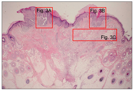
Fig. 2 Histopathology of the yellow, verrucous plaque. Hyperkeratosis, irregular acanthosis, and papillomatosis were present in the epidermis. Incompletely differentiated hair structures were in the dermis (H&E, ×12.5).

Fig. 3 (A) Cystic invagination extended downward from the epidermis (H&E, ×40), which was diagnosed as a syringocystadenoma papilliferum. (B) There was a parakeratotic column overlying the trichoblastoma like-lesion, resembling the cornoid lamella of porokeratosis (H&E, ×40). (C) There were fat cells in upper dermis (H&E, ×40). (D) The basaloid epithelial proliferation showed PAS negative finding (PAS, ×100).
Reference
-
1. Mehregan AH, Pinkus H. Life history of organoid nevi. Special reference to nevus sebaceous of Jadassohn. Arch Dermatol. 1965. 91:574–588.2. Baker BB, Imber RJ, Templer JW. Nevus sebaceous of Jadassohn. Arch Otolaryngol. 1975. 101:515–516.
Article3. Cribier B, Scrivener Y, Grosshans E. Tumors arising in nevus sebaceus: a study of 596 cases. J Am Acad Dermatol. 2000. 42:263–268.4. Jaqueti G, Requena L, Sánchez Yus E. Trichoblastoma is the most common neoplasm developed in nevus sebaceus of Jadassohn: a clinicopathologic study of a series of 155 cases. Am J Dermatopathol. 2000. 22:108–118.
Article5. Jones EW, Heyl T. Naevus sebaceus. A report of 140 cases with special regard to the development of secondary malignant tumours. Br J Dermatol. 1970. 82:99–117.6. Stavrianeas NG, Katoulis AC, Stratigeas NP, Karagianni IN, Patertou-Stavrianea M, Varelzidis AG. Development of multiple tumors in a sebaceous nevus of Jadassohn. Dermatology. 1997. 195:155–158.
Article7. Nakai K, Yoneda K, Moriue J, Moriue T, Matsuoka Y, Kubota Y. Sebaceoma, trichoblastoma and syringocystadenoma papilliferum arising within a nevus sebaceous. J Dermatol. 2008. 35:365–367.
Article8. Maize JC, Foster G. Age-related changes in melanocytic naevi. Clin Exp Dermatol. 1979. 4:49–58.
Article9. Ahn SK, Ahn HJ, Kim TH, Hwang SM, Choi EH, Lee SH. Intratumoral fat in neurofibroma. Am J Dermatopathol. 2002. 24:326–329.
Article10. Reed RJ, Leone P. Porokeratosis: a mutant clonal keratosis of the epidermis. I. Histogenesis. Arch Dermatol. 1970. 101:340–347.
Article
- Full Text Links
-
- Actions
-
Cited
- CITED
-
- Close
- Share
-
- Similar articles
-
- Eccrine Poroma Arising within Nevus Sebaceous
- Development of seven secondary neoplasms in a nevus sebaceous: a case report and literature review
- A case of sebaceous carcinoma arising from nevus sebaceus of jadassohn
- Sebaceous Carcinoma Arising from Nevus Sebaceus
- A Case of Sebaceous Epithelioma Arising from Nevus Sebaceus
