Late-Onset Eccrine Angiomatous Hamartoma Associated with a Ganglion Cyst on the Sole of the Foot
- Affiliations
-
- 1Department of Dermatology and Institute of Hair and Cosmetic Medicine, Yonsei University Wonju College of Medicine, Wonju, Korea. leewonsoo@yonsei.ac.kr
- KMID: 2156794
- DOI: http://doi.org/10.5021/ad.2011.23.S2.S218
Abstract
- Eccrine angiomatous hamartoma (EAH) is a benign, uncommon, combined vascular and eccrine malformation. Most cases of this disorder have been single or multiple nodules or plaques that appear red, yellow, blue, violaceous, or skin colored. EAH may be congenital or appear later in childhood; it rarely arises during puberty or adulthood. A 52-year-old female patient visited our department for tender subcutaneous cystic tumor on the right sole with a one month history. Histopathologic examination confirmed EAH. During excisional biopsy procedure, mucinous discharges were observed which were histopathologically diagnosed as ganglion.
Keyword
Figure
-
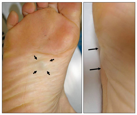
Fig. 1 The patient presented with a solitary subcutaneous tumor with intact epidermis on the sole of the right foot.

Fig. 2 Histopathologic examination of the lesion showed increased numbers of eccrine glands, as well as dilated vascular channels in the deep dermis and subcutaneous tissue (H&E, ×12.5).

Fig. 3 Increased numbers of eccrine glands were associated with dilated vascular channels in the deep dermis and subcutaneous tissue (H&E, ×40, ×100).
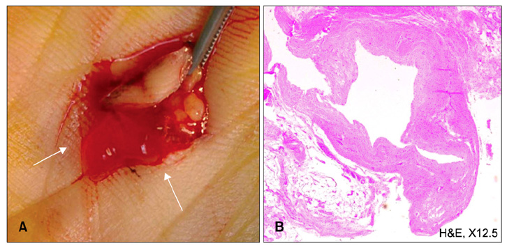
Fig. 4 During excisional biopsy, gelatinous material (A) was noted and later confirmed as a ganglion cyst by histopathologic findings, including a cystic space surrounded with collagenous fibers instead of epithelial lining (B: H&E, ×12.5).
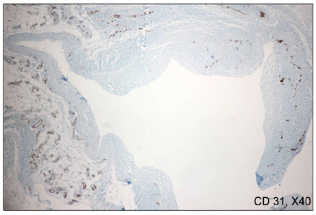
Fig. 5 Specific staining with CD 31 revealed no endothelial lining in cystic space (CD 31, ×40).
Cited by 1 articles
-
Eccrine Angiomatous Hamartoma: A Review of Ten Cases
Jaeyoung Shin, Yong Hyun Jang, Soo-Chan Kim, You Chan Kim
Ann Dermatol. 2013;25(2):208-212. doi: 10.5021/ad.2013.25.2.208.
Reference
-
1. Lotzbeck C. Ein Fall von Schweissdrsengeschwulst an der Wauge. Virchow Arch Pathol Anat Physiol Klin Med. 1859. 16:160.2. Hyman AB, Harris H, Brownstein MH. Eccrine angiomatous hamartoma. N Y State J Med. 1968. 68:2803–2806.3. Pelle MT, Pride HB, Tyler WB. Eccrine angiomatous hamartoma. J Am Acad Dermatol. 2002. 47:429–435.
Article4. Martinelli PT, Tschen JA. Eccrine angiomatous hamartoma: a case report and review of the literature. Cutis. 2003. 71:449–455.5. Zeller DJ, Goldman RL. Eccrine-pilar angiomatous hamartoma. Report of a unique case. Dermatologica. 1971. 143:100–104.6. Campen R, Zembowicz A, Liu V, Wrone D. Linear ectodermal cutaneous hamartoma. Int J Dermatol. 2003. 42:376–379.
Article7. Castilla EA, Schwimer CJ, Bergfeld WF, Skacel M, Ormsby A. Eccrine angiomatous hamartoma in a neurofibromatosis type-1 patient. Pathology. 2002. 34:378–380.
Article8. Donati P, Amantea A, Balus L. Eccrine angiomatous hamartoma: a lipomatous variant. J Cutan Pathol. 1989. 16:227–229.
Article9. Enjolras O, Mulliken JB. Vascular tumors and vascular malformations (new issues). Adv Dermatol. 1997. 13:375–423.10. Seraly MP, Magee K, Abell E, Bridenstine J, Jegasothy BV. Eccrine-angiomatous nevus, a new variant. J Am Acad Dermatol. 1993. 29:274–275.
Article11. Sulica RL, Kao GF, Sulica VI, Penneys NS. Eccrine angiomatous hamartoma (nevus): immunohistochemical findings and review of the literature. J Cutan Pathol. 1994. 21:71–75.
Article12. Kirby EJ, Shereff MJ, Lewis MM. Soft-tissue tumors and tumor-like lesions of the foot. An analysis of eighty-three cases. J Bone Joint Surg Am. 1989. 71:621–626.
Article13. Soren A. Pathogenesis and treatment of ganglion. Clin Orthop Relat Res. 1966. 48:173–179.14. Psaila JV, Mansel RE. The surface ultrastructure of ganglia. J Bone Joint Surg Br. 1978. 60:228–233.
Article15. Chloros GD, Wiesler ER, Poehling GG. Current concepts in wrist arthroscopy. Arthroscopy. 2008. 24:343–354.
Article16. Plate AM, Lee SJ, Steiner G, Posner MA. Tumorlike lesions and benign tumors of the hand and wrist. J Am Acad Orthop Surg. 2003. 11:129–141.
Article17. Pontious J, Good J, Maxian SH. Ganglions of the foot and ankle. A retrospective analysis of 63 procedures. J Am Podiatr Med Assoc. 1999. 89:163–168.
Article18. Wolf R, Krakowski A, Dorfman B, Baratz M. Eccrine angiomatous hamartoma. A painful step. Arch Dermatol. 1989. 125:1489–1490.
Article19. Gabrielsen TO, Elgjo K, Sommerschild H. Eccrine angiomatous hamartoma of the finger leading to amputation. Clin Exp Dermatol. 1991. 16:44–45.
Article20. Nakayama H, Mihara M, Hattori K, Mishima E, Shimao S. Eccrine angiomatous hamartoma of the sacral region. Acta Derm Venereol. 1994. 74:477.21. Cebreiro C, Sánchez-Aguilar D, Gómez Centeno P, Fernández-Redondo V, Toribio J. Eccrine angiomatous hamartoma: report of seven cases. Clin Exp Dermatol. 1998. 23:267–270.
Article22. Tsuji T, Sawada H. Eccrine angiomatous hamartoma with verrucous features. Br J Dermatol. 1999. 141:167–169.
Article23. Laeng RH, Heilbrunner J, Itin PH. Late-onset eccrine angiomatous hamartoma: clinical, histological and imaging findings. Dermatology. 2001. 203:70–74.
Article24. Jeong E, Park HJ, Oh ST, Lee JY, Cho BK. Late-onset eccrine angiomatous hamartoma on the forehead. Int J Dermatol. 2006. 45:598–599.
Article
