Ann Dermatol.
2011 May;23(2):222-224. 10.5021/ad.2011.23.2.222.
Linear Lichen Sclerosus along the Blaschko's Line of the Face
- Affiliations
-
- 1Department of Dermatology, Samsung Medical Center, Sungkyunkwan University School of Medicine, Seoul, Korea. dylee@skku.edu
- KMID: 2156668
- DOI: http://doi.org/10.5021/ad.2011.23.2.222
Abstract
- Lichen sclerosus et atrophicus (LSA) is an inflammatory disease that primarily causes anogenital lesion in middle aged women. We present here a case of facial LSA with an asymptomatic, well-demarcated, whitish to bluish, atrophic patch in a linear pattern on the forehead of a 48-year-old woman. This case showed an atypical clinical presentation and it mimicked en coup de sabre, but the histopathologic results confirmed the diagnosis of LSA.
MeSH Terms
Figure
-
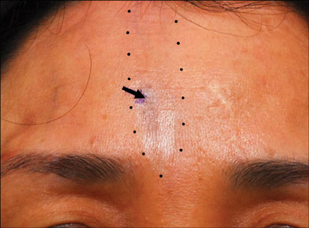
Fig. 1 A linear, well-demarcated atrophic patch on the forehead (black arrow).
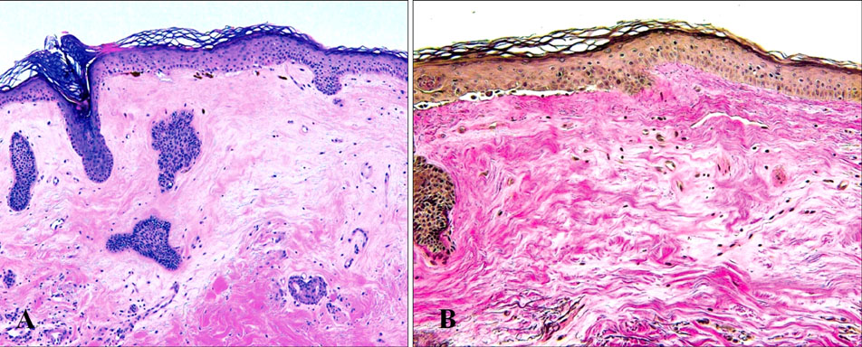
Fig. 2 (A) Thinning of the epidermis, loss of the rete ridges, focal basal cell vacuolization, pigmentary incontinence, edema and hyalination of the papillary dermis along with a moderate lymphomononuclear cell infiltrate are noted (H&E stain, ×100). (B) Special staining for elastic fiber showed scanty elastic tissue in the dermis (Elastic fiber, ×100).
Reference
-
1. Meffert JJ, Davis BM, Grimwood RE. Lichen sclerosus. J Am Acad Dermatol. 1995. 32:393–416.
Article2. Izumi T, Tajima S. A case of linear type of lichen sclerosus et atrophicus? J Dermatol. 1995. 22:279–282.
Article3. Kim YJ, Lee ES. Case of sequentially occurring lesions of facial lichen sclerosus following the lines of Blaschko. J Dermatol. 2007. 34:201–204.
Article4. Kaur S, Thami GP, Kanwar AJ, Mohan H. Linear oro-facial lichen sclerosus. Clin Exp Dermatol. 2002. 27:467–470.
Article5. Walsh SN, Jorizzo JL, Haverstock C, Sangüeza OP. A linear orofacial macule. Am J Dermatopathol. 2008. 30:194–195.
Article6. Happle R, Assim A. The lines of Blaschko on the head and neck. J Am Acad Dermatol. 2001. 44:612–615.
Article7. Hengge UR. Wolff K, Goldsmith LA, Katz SI, Gilchrest BA, Paller AS, Leffell DJ, editors. Lichen sclerosus. Fitzpatrick's dermatology in general medicine. 2008. 7th ed. New York: McGraw-Hill;546–550.8. McNiff JM, Glusac EJ, Lazova RZ, Carroll CB. Morphea limited to the superficial reticular dermis: an underrecognized histologic phenomenon. Am J Dermatopathol. 1999. 21:315–319.
Article9. Tremaine R, Adam JE, Orizaga M. Morphea coexisting with lichen sclerosus et atrophicus. Int J Dermatol. 1990. 29:486–489.
Article10. Sawamura D, Yaguchi T, Hashimoto I, Nomura K, Konta R, Umeki K. Coexistence of generalized morphea with hisotological changes in lichen sclerosus et atrophicus and lichen planus. J Dermatol. 1998. 25:409–411.
Article11. Nishioka S. Histological comparison of morphea and lichen sclerosus et atrophicus. Kurume Med J. 1997. 44:83–90.
Article12. Kreuter A, Gambichler T, Avermaete A, Happe M, Bacharach-Buhles M, Hoffmann K, et al. Low-dose ultraviolet A1 phototherapy for extragenital lichen sclerosus: results of a preliminary study. J Am Acad Dermatol. 2002. 46:251–255.
Article13. Assmann T, Becker-Wegerich P, Grewe M, Megahed M, Ruzicka T. Tacrolimus ointment for the treatment of vulvar lichen sclerosus. J Am Acad Dermatol. 2003. 48:935–937.
Article
