Ann Dermatol.
2008 Dec;20(4):237-239. 10.5021/ad.2008.20.4.237.
Development of Halo Nevus Around Nevus Spilus as a Central Nevus, and the Concurrent Vitiligo
- Affiliations
-
- 1Department of Dermatology, Kangnam St. Mary's Hospital, College of Medicine, The Catholic University of Korea, Seoul, Korea. yykim@catholic.ac.kr
- KMID: 2156403
- DOI: http://doi.org/10.5021/ad.2008.20.4.237
Abstract
- Halo nevus is a benign melanocytic nevus that is surrounded by a hypopigmented zone. The most frequent association with halo nevus is vitiligo, and this also appears in nearby regions, as well as at other remote sites. Although the mechanism for developing the depigmentation around nevus spilus is uncertain an immunologic process may be responsible for the finding of inflammatory infiltrates of the upper dermis in the depigmented lesions. We report here on a 13-year-old boy who showed a depigmented zone around a nevus spilus on the right side of his neck with simultaneous vitiligo lesions on the face.
Keyword
Figure
-
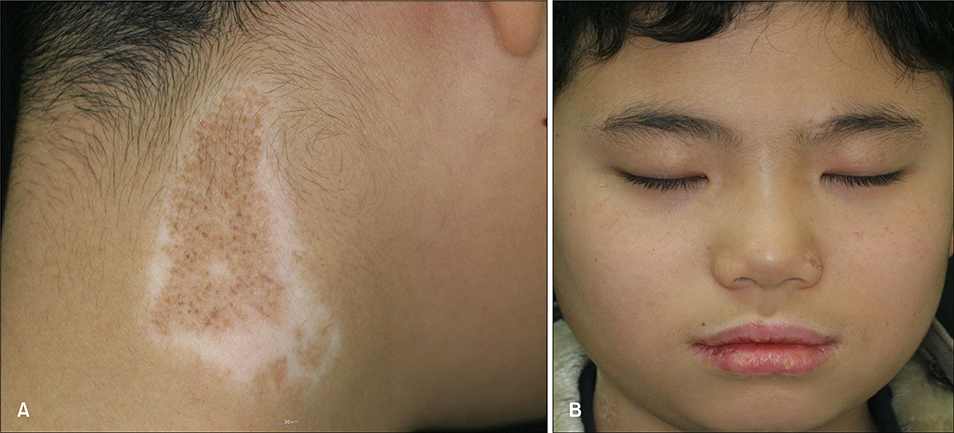
Fig. 1 (A) A well demarcated, pale brown colored patch with multiple, scattered, dark brown speckles on the right side of the neck with a hypopigmented zone around the nevus. (B) In addition, lesions of vitiligo were found in the perioral, perinasal and periorbital regions.
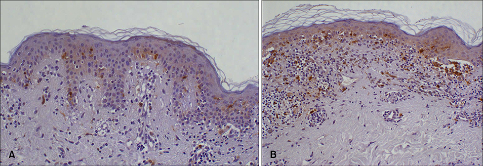
Fig. 2 (A) The histopathology of the pale brown colored patch lesion revealed elongation of the rete ridges and increased melanin pigmentation in the basal layer of the epidermis. (B) Biopsy from the dark brown colored speckles showed increased melanin in the dermoepidermal junction and upper dermis. Mononuclear cells and lymphohistiocytes were infiltrated into the upper part of the dermis in both lesions (A, B) (S-100, ×100).
Reference
-
1. Langer K, Konrad K. Congenital melanocytic nevi with halo phenomenon: report of two cases and a review of the literature. J Dermatol Surg Oncol. 1990; 16:377–380.
Article2. Itin PH, Lautenschlager S. Acquired leukoderma in congenital pigmented nevus associated with vitiligo-like depigmentation. Pediatr Dermatol. 2002; 19:73–75.
Article3. Guerra-Tapia A, Isarria MJ. Periocular vitiligo with onset around a congenital divided nevus of the eyelid. Pediatr Dermatol. 2005; 22:427–429.
Article4. Brandt O, Christophers E, Folster-Holst R. Halo dermatitis followed by the development of vitiligo associated with Sutton's nevi. J Am Acad Dermatol. 2005; 52:5 Suppl 1. S101–S104.
Article5. Zeff RA, Freitag A, Grin CM, Grant-Kels JM. The immune response in halo nevi. J Am Acad Dermatol. 1997; 37:620–624.
Article6. Reed RJ, Ichinose H, Clark WH Jr, Mihm MC Jr. Common and uncommon melanocytic nevi and borderline melanomas. Semin Oncol. 1975; 2:119–147.7. Mooney MA, Barr RJ, Buxton MG. Halo nevus or halo phenomenon? A study of 142 cases. J Cutan Pathol. 1995; 22:342–348.
Article8. Schallreuter KU, Kothari S, Elwary S, Rokos H, Hasse S, Panske A. Molecular evidence that halo in Sutton's naevus is not vitiligo. Arch Dermatol Res. 2003; 295:223–228.
Article9. de Vijlder HC, Westerhof W, Schreuder GM, de Lange P, Claas FH. Difference in pathogenesis between vitiligo vulgaris and halo nevi associated with vitiligo is supported by an HLA association study. Pigment Cell Res. 2004; 17:270–274.
Article10. Shin JH, Kim MJ, Cho S, Whang KK, Hahm JH. A case of giant congenital nevocytic nevus with neurotization and onset of vitiligo. J Eur Acad Dermatol Venereol. 2002; 16:384–386.
Article
