Ann Dermatol.
2008 Dec;20(4):197-199. 10.5021/ad.2008.20.4.197.
A Case of Intradermal Melanocytic Nevus with Ossification (Nevus of Nanta)
- Affiliations
-
- 1Department of Dermatology, College of Medicine, The Catholic University of Korea, Seoul, Korea. cjpark777@yahoo.co.kr
- KMID: 2156393
- DOI: http://doi.org/10.5021/ad.2008.20.4.197
Abstract
- A 49-year-old woman presented with a 30-year history of asymptomatic plaque on her right temple. The histological examination revealed nests of nevus cells throughout the entire dermis. Bony spicules were seen just beneath the nevus cell nests in the lower dermis. Cutaneous ossification is an unusual event. Herein, we present a case of intradermal melanocytic nevus with unusual ossification (nevus of Nanta). To the best of our knowledge, this is the first such case report in the Korean literature.
Keyword
Figure
-

Fig. 1 A 1.5 cm in diameter, slightly erythematous to flesh colored, flat-topped hairy plaque on the right temple of the face.
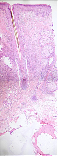
Fig. 2 Nests of nevus cells were seen within the upper dermis, and bony spicules surrounding the mature fatty tissue were observed in the lower dermis, underneath the nevus cell nests (Hematoxylin-eosin, ×40).

Fig. 3 Bony spicules containing numerous osteocytes(Hematoxylin-eosin, ×100).
Reference
-
1. Burgdorf W, Nasemann T. Cutaneous osteomas:a clinical and histopathologic review. Arch Der-mato Res. 1977; 260:121–135.2. Elder DE, Elenitsas R, Johnson BL Jr, Murphy GF. Lever's Histopathology of the skin. 19th ed. Philadelphia: Lippincott Williams & Wilkins;2005. p. 1089–1093.3. Moulin G, Souquet D, Balme B. Pigmented nevus and cutaneous ossifications. Apropos of 125 cases of osteonevi. Ann Dermatol Venereol. 1991; 118:199–204.4. Kanitakis J, Claudy A. Mummified ossified melanocytic naevus. Eur J Dermatol. 2000; 10:466–467.5. Orlow SJ, Watsky KL, Bolognia JL. Skin and bones. II. J Am Acad Dermatol. 1991; 25:447–462.
Article6. Conlin PA, Jimenez-Quintero LP, Rapini RP. Osteomas of the skin revisited: a clinicopathologic review of 74 cases. Am J Dermatopathol. 2002; 24:479–483.7. Sasaki S, Mitsuhashi Y, Ito Y. Osteo-nevus of Nanta: a case report and review of the Japanese literature. J Dermatol. 1999; 26:183–188.
Article8. Keida T, Hayashi N, Kawakami M, Kawashima M. Transforming growth factor beta and connective tissue growth factor are involved in the evolution of nevus of Nanta. J Dermatol. 2005; 32:442–445.
Article9. Culver W, Burgdorf WH. Malignant melanoma arising in a nevus of Nanta. J Cutan Pathol. 1993; 20:375–377.
Article
