Unusual Cause of Hip Pain: Intrusion of the Acetabular Labrum
- Affiliations
-
- 1Department of Orthopaedic Surgery, Daegu Fatima Hospital, Daegu, Korea. femur1973@hanmail.net
- KMID: 2156020
- DOI: http://doi.org/10.5371/hp.2015.27.1.49
Abstract
- Femoroacetabular impingement and dysplatic hip joint is well known cause of osteoarthritis. In these diseases, labral tear and subsequent cartilage damage is thought to be main pathophysiology of development of osteoarthritis. If there are no known bony abnormalities, we called it as idiopathic osteoarthritis. Normal appearance of acetabular labrum is a continuous, usually triangular structure that attaches to the bony rim of the acetabulum and is completed at the inferior portion by the transverse acetabular ligament over the acetabular notch. A few authors reported intra-articular labrum and its relation to the development of osteoarthritis. But they didn't comment the primary bony abnormality especially acetabulum. We'd like to report x-ray, computed tomogram, magnetic resonance arthrogram and arthroscopic findings of a case had double contour sign of acetabular dome combined with intrusion of acetabular labrum.
Keyword
Figure
-
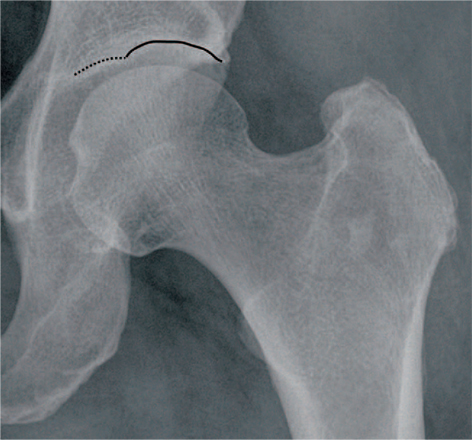
Fig. 1 Left hip anteroposterior view shows joint space discrepancy between the medial (dotted line) and lateral acetabular dome (solid line) like gull wing. It means there is two distinct contour at the acetabular dome.
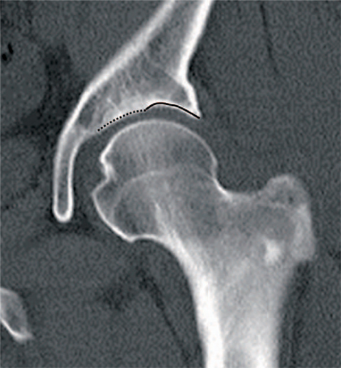
Fig. 2 Definite double contour (dotted and solid line) at the acetabular dome can be seen at the coronal reconstruction computed tomogram scanning.
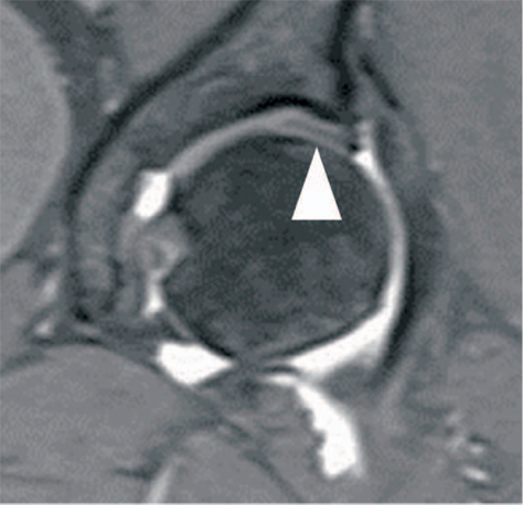
Fig. 3 Coronal reconstruction of magnetic resonance arthrogram shows intra-articular low signal intensity line indicating intrusion of acetabular labrum (arrowhead).
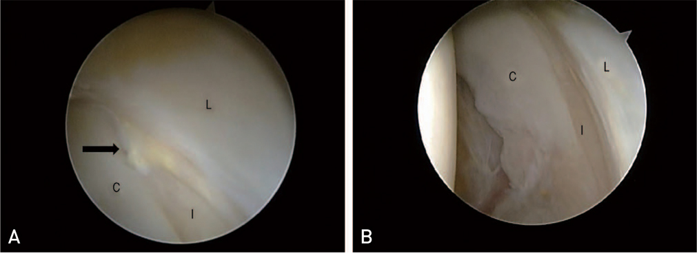
Fig. 4 (A) Arthroscopic picture viewing from anterolateral portal shows torn cartilage (arrow), acetabular labrum (L), acetabular cartilage (C), intrusion of acetabular labrum (I). (B) After shaving and cauterization of the torn cartilage, reduced acetabular cartilage width from the acetabular fossa is shown clearly.
Reference
-
1. Milgram JW, Tachdjian MO. Pathology of the limbus in untreated teratologic congenital dislocation of the hip. A case report of a ten-month-old- infant. Clin Orthop Relat Res. 1976; 119:107–111.
Article2. Harris WH, Bourne RB, Oh I. Intra-articular acetabular labrum: a possible etiological factor in certain cases of osteoarthritis of the hip. J Bone Joint Surg Am. 1979; 61:510–514.3. Byrd JW, Jones KS. Osteoarthritis caused by an inverted acetabular labrum: radiographic diagnosis and arthroscopic treatment. Arthroscopy. 2002; 18:741–747.4. Guevara CJ, Pietrobon R, Carothers JT, Olson SA, Vail TP. Comprehensive morphologic evaluation of the hip in patients with symptomatic labral tear. Clin Orthop Relat Res. 2006; 453:277–285.
Article5. Peelle MW, Della Rocca GJ, Maloney WJ, Curry MC, Clohisy JC. Acetabular and femoral radiographic abnormalities associated with labral tears. Clin Orthop Relat Res. 2005; 441:327–333.
Article6. Ferguson SJ, Bryant JT, Ganz R, Ito K. An in vitro investigation of the acetabular labral seal in hip joint mechanics. J Biomech. 2003; 36:171–178.
Article7. Altenberg AR. Acetabular labrum tears: a cause of hip pain and degenerative arthritis. South Med J. 1977; 70:174–175.8. Kim YH. Acetabular dysplasia and osteoarthritis developed by an eversion of the acetabular labrum. Clin Orthop Relat Res. 1987; (215):289–295.
Article9. Ganz R, Leunig M, Leunig-Ganz K, Harris WH. The etiology of osteoarthritis of the hip: an integrated mechanical concept. Clin Orthop Relat Res. 2008; 466:264–272.
- Full Text Links
-
- Actions
-
Cited
- CITED
-
- Close
- Share
-
- Similar articles
-
- Acetabular Dysplasia and Osteoarthritis Developed by an Eversion of the Acetabular Labrum
- Direct MR Arthrography of the Hip: Diagnosis and Pitfalls of Acetabular Labral Lesions
- Hip Arthroscopy for Incarcerated Acetabular Labrum following Reduction of Traumatic Hip Dislocation: Three Case Reports
- Changes of the hip joints associated with chronic subluxation and dislocation: CT and plain radiographic analysis
- Age-related Morphologic and Pathologic Features of Acetabular Labrum in the Adult Hip
