Korean J Leg Med.
2016 Feb;40(1):27-31. 10.7580/kjlm.2016.40.1.27.
Mixture Patterned Short Tandem Repeat Profile in a Perimortem Transfused Patient
- Affiliations
-
- 1Department of Forensic Medicine, Seoul National University College of Medicine, Seoul, Korea. sdlee@snu.ac.kr
- 2Institute of Forensic Science, Seoul National University College of Medicine, Seoul, Korea.
- 3Medical Examiner's Office, National Forensic Service, Wonju, Korea.
- KMID: 2155728
- DOI: http://doi.org/10.7580/kjlm.2016.40.1.27
Abstract
- Recently, it has been reported that transfused patients can generate admixture-like genetic profiles. As genetic material of the donor can survive for a reasonable time after transfusion, the recipient's genomic DNA is likely to have a mixture pattern. An autopsy case of a man transfused perimortem generated a mixture patterned short tandem repeat profile. Notably, the patient was transfused mostly with nuclear-deficient cells, limiting the donor genetic material available for the recipient. As a result, mixture-like patterns were observed consistently, regardless of change in input DNA content; the sample DNA content, which was serially diluted, ranged from 1 ng to 0.0625 ng. The distributions of foreign peaks appeared to be irreproducible, showing stochastic behaviors throughout the genotyped results. This study suggests that a cautious approach is required when genotyping of a patient who has undergone recent transfusion. One must consider the possibility of obtaining a mixture patterned profile in such patients, and therefore, choose parenchymal organs or tissues for reliable results.
MeSH Terms
Figure
-
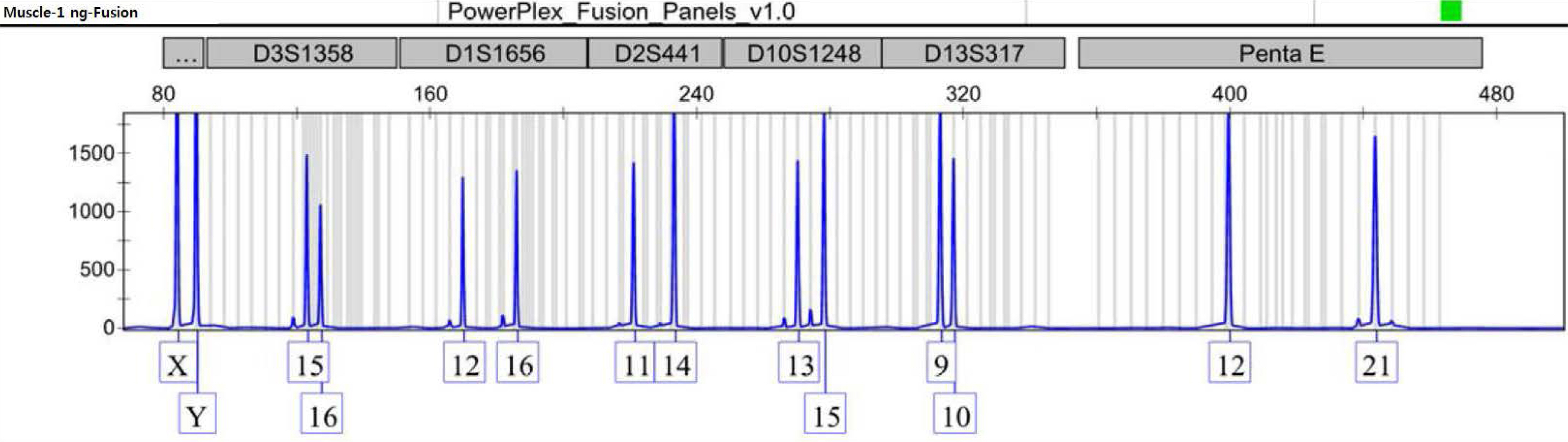
Fig. 1. An example of the patient's consensus profile from a muscle tissue sample.

Fig. 2. Overview of foreign peaks detected in blood DNA sample diluted to 0.5 ng (A) and 0.125 ng (B).
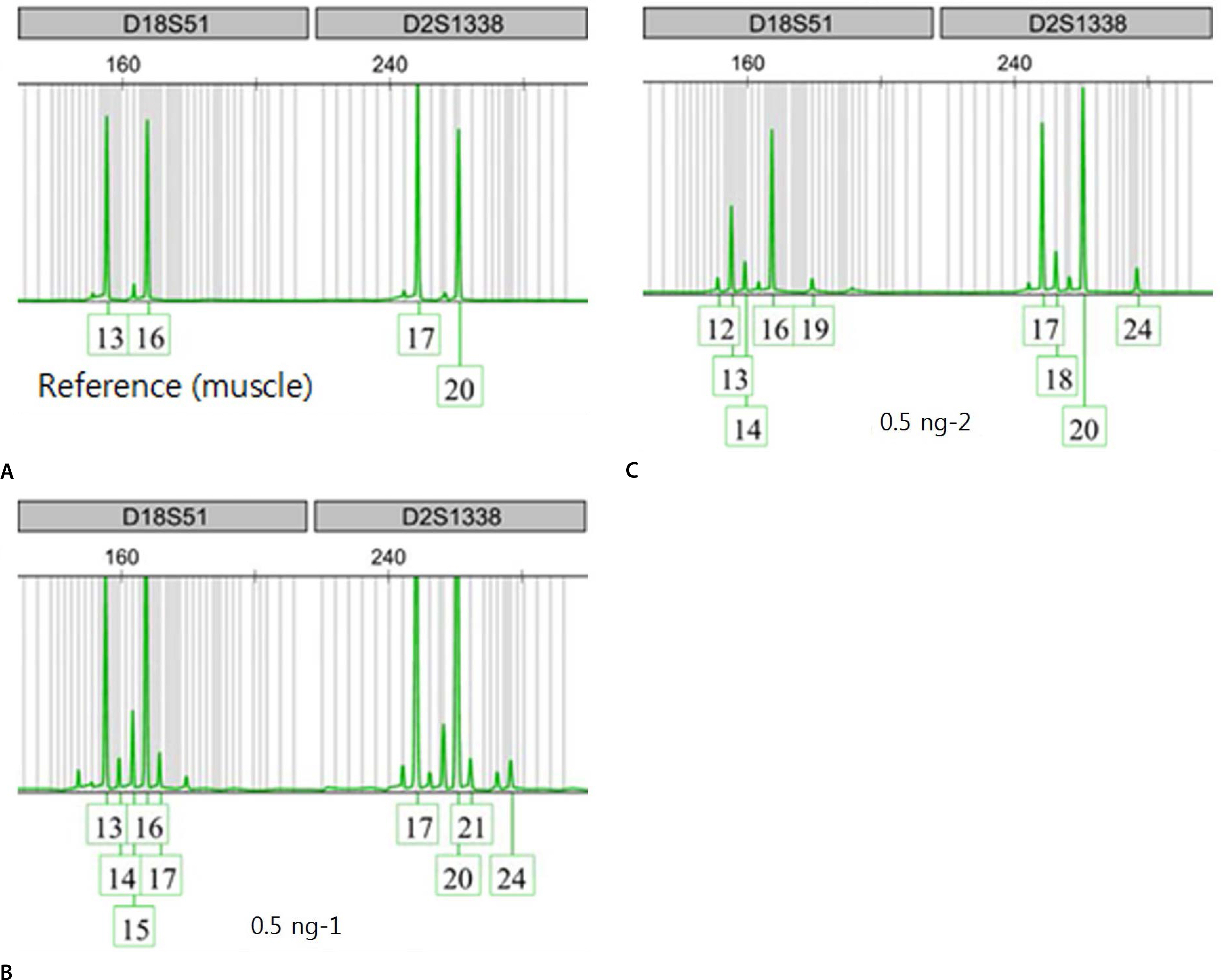
Fig. 3. An example of alleles at two loci obtained from 1 ng of muscle DNA sample as reference (A), and 0.5 ng of blood DNA samples in duplicates (B, C).
Reference
-
1.Wenk RE., Chiafari PA. DNA typing of recipient blood after massive transfusion. Transfusion. 1997. 37:1108–10.
Article2.Graham EA., Tsokos M., Rutty GN. Can post-mortem blood be used for DNA profiling after peri-mortem blood transfusion? Int J Legal Med. 2007. 121:18–23.
Article3.Dauber EM., Dorner G., Mitterbauer M, et al. Discrepant results of samples taken from different tissues of a single individual. Int Congr Ser. 2004. 1261:48–9.
Article4.Vietor HE., Hallensleben E., van Bree SP, et al. Survival of donor cells 25 years after intrauterine transfusion. Blood. 2000. 95:2709–14.5.Gong MN., Sai Y., Zhou W, et al. Genotyping patients with recent blood transfusions. Epidemiology. 2003. 14:744–7.
Article6.Lee TH., Montalvo L., Chrebtow V, et al. Quantitation of genomic DNA in plasma and serum samples: higher concentrations of genomic DNA found in serum than in plasma. Transfusion. 2001. 41:276–82.
Article7.Schechter GP., Whang-Peng J., McFarland W. Circulation of donor lymphocytes after blood transfusion in man. Blood. 1977. 49:651–6.
Article8.Utter GH., Reed WF., Lee TH, et al. Transfusion-associated microchimerism. Vox Sang. 2007. 93:188–95.
Article9.Jung JY., Park SH., Park SW, et al. Mixed DNA profiles in transfused deceased case. Korean J Sci Crim Invest. 2015. 9:206–12.
- Full Text Links
-
- Actions
-
Cited
- CITED
-
- Close
- Share
-
- Similar articles
-
- Rapid Prenatal Detection of Down and Edwards Syndromes by Fluorescent Polymerase Chain Reaction with Short Tandem Repeat Markers
- Sequence Generation and Genotyping of 15 Autosomal STR Markers Using Next Generation Sequencing
- DNA Profiling via Short Tandem Repeat Analysis by Using Serum Samples
- Instability at Short Tandem Repeats in Lymphoblastoid Cell Lines
- Comparison of the Effects of Patterned and Conventional Panretinal Photocoagulation on Diabetic Retinopathy