Computed Tomography and Magnetic Resonance Imaging Findings of Nasal Cavity Hemangiomas According to Histological Type
- Affiliations
-
- 1Department of Radiology, Inha University School of Medicine, Incheon 400-711, Korea. swpark8802@gmail.com
- 2Department of Radiology, Seoul National University College of Medicine, Seoul 110-744, Korea.
- 3Department of Radiology, Boramae Medical Center, Seoul 156-707, Korea.
- 4Department of Otolaryngology-Head & Neck Surgery, Inha University School of Medicine, Incheon 400-711, Korea.
- 5Department of Radiology, Inha University Hospital, Incheon 400-711, Korea.
- KMID: 2155526
- DOI: http://doi.org/10.3348/kjr.2015.16.3.566
Abstract
OBJECTIVE
To compare computed tomography (CT) and magnetic resonance imaging (MRI) findings between two histological types of nasal hemangiomas (cavernous hemangioma and capillary or lobular capillary hemangioma).
MATERIALS AND METHODS
CT (n = 20; six pre-contrast; 20 post-enhancement) and MRI (n = 7) images from 23 patients (16 men and seven women; mean age, 43 years; range, 13-73 years) with a pathologically diagnosed nasal cavity hemangioma (17 capillary and lobular capillary hemangiomas and six cavernous hemangiomas) were reviewed, focusing on lesion location, size, origin, contour, enhancement pattern, attenuation or signal intensity (SI), and bony changes.
RESULTS
The 17 capillary and lobular hemangiomas averaged 13 mm (range, 4-37 mm) in size, and most (n = 13) were round. Fourteen capillary hemangiomas had marked or moderate early phase enhancement on CT, which dissipated during the delayed phase. Four capillary hemangiomas on MRI showed marked enhancement. Bony changes were usually not seen on CT or MRI (seen on five cases, 29.4%). Half of the lesions (2/4) had low SI on T1-weighted MRI images and heterogeneously high SI with signal voids on T2-weighted images. The six cavernous hemangiomas were larger than the capillary type (mean, 20.5 mm; range, 10-39 mm) and most had lobulating contours (n = 4), with characteristic enhancement patterns (three centripetal and three multifocal nodular), bony remodeling (n = 4, 66.7%), and mild to moderate heterogeneous enhancement during the early and delayed phases.
CONCLUSION
CT and MRI findings are different between the two histological types of nasal hemangiomas, particularly in the enhancement pattern and size, which can assist in preoperative diagnosis and planning of surgical tumor excision.
Keyword
MeSH Terms
Figure
-
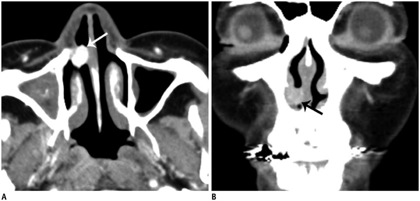
Fig. 1 Computed tomography (CT) image of 59-year-old woman who presented with nasal congestion. Right nasal tumor was diagnosed as lobular capillary hemangioma. A. Axial early-phase enhanced CT image reveals well-defined round mass with marked enhancement in right anterior nasal cavity (white arrow). B. Coronal delayed-phase enhanced CT image shows dissipation of tumor enhancement. Tumor reveals mild enhancement without adjacent bony changes (black arrow).
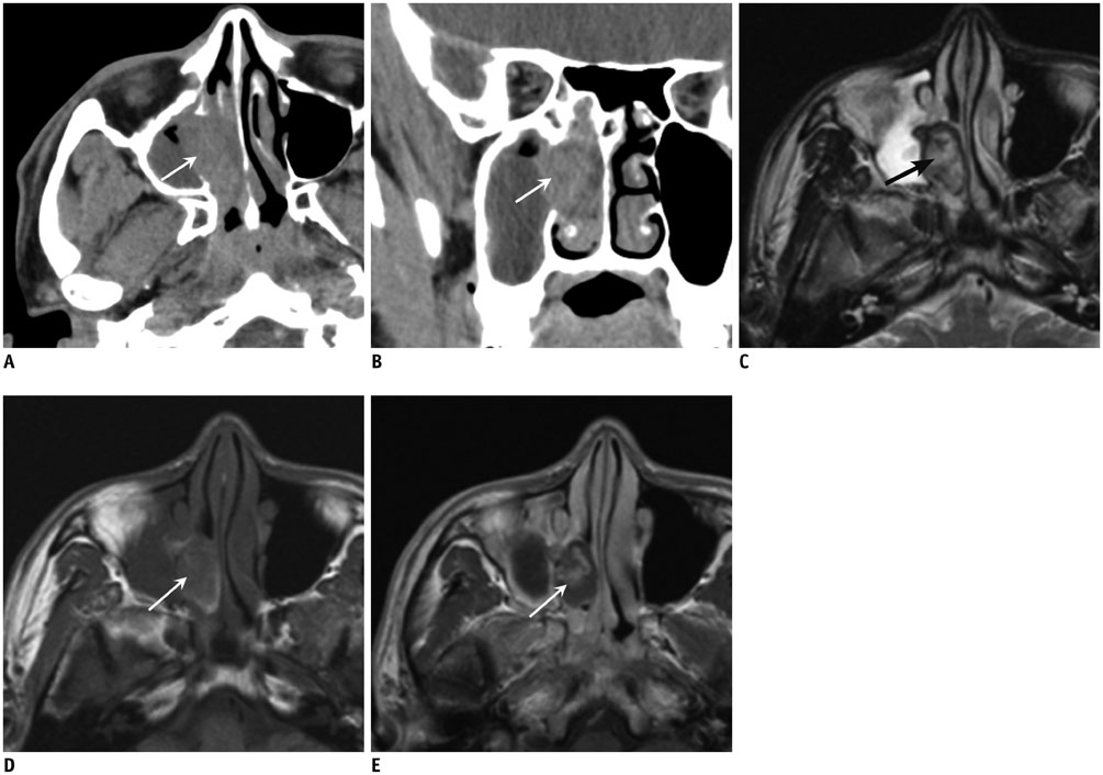
Fig. 2 Computed tomography (CT) and magnetic resonance images of 34-year-old man with right nasal tumor diagnosed as cavernous hemangioma. A. Early-phase CT image shows mild heterogeneous enhancing tumor (arrow) in right middle meatus. B. Tumor has mild heterogeneous enhancement on coronal delayed-phase image (arrow). Coronal CT image shows erosion of right middle turbinate and lateral nasal wall. C. Mass has heterogeneous high and low signal intensity on T2-weighted axial image (arrow). D. Mass appears with iso-signal intensity (arrow) on pre-contrast T1-weighted axial image. High signal intensity area on tumor margin of T1-weighted image suggests thrombus or thick mucus retention. E. Mass has partial heterogeneous mild enhancement (arrow) on post-contrast T1-weighted axial image.
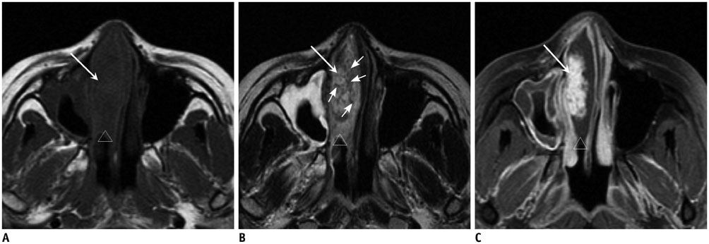
Fig. 3 Magnetic resonance imaging (MRI) of 31-year-old woman who presented with recurrent right nasal hemorrhage and nasal congestion. Nasal tumor was histologically diagnosed as capillary hemangioma. A. Axial T1-weighted image reveals lobulating contoured mass in right anterior nasal cavity and middle meatus (white arrow). Mass has low signal intensity on T1-weighted image. Peritumoral cystic region in tumor periphery has low signal intensity on T1-weighted image (empty arrowhead). B. Right nasal mass has heterogeneously high signal intensity (large arrow) on axial T2-weighted image. There are irregular signal voids within mass (small arrows). Peritumoral cystic region has high signal intensity on T2-weighted image (empty arrowhead). C. Axial T1-weighted contrast-enhanced MRI shows markedly enhanced mass (white arrow) displacing right middle turbinate and deviating nasal septum leftward. Peritumoral cystic region exhibits no contrast enhancement (empty arrowhead).
Cited by 2 articles
-
Extranasopharyngeal Angiofibroma of the Nasal Septum: A Case Report
Gyoung-Eun Lee, Tae Gyu Kim, Kyung-Eun Bae, Kyoung Rai Cho, Jung Heob Sohn, Bo Young Kim, Hyun-jung Kim, Guhyun Kang
J Korean Soc Radiol. 2019;80(4):750-755. doi: 10.3348/jksr.2019.80.4.750.A Case of 2-Month-Old Infant with Lobular Capillary Hemangioma
Yong Seok Kang, Young Kang, Doo Hee Han
J Rhinol. 2017;24(2):127-131. doi: 10.18787/jr.2017.24.2.127.
Reference
-
1. Dillon WP, Som PM, Rosenau W. Hemangioma of the nasal vault: MR and CT features. Radiology. 1991; 180:761–765.2. Iwata N, Hattori K, Nakagawa T, Tsujimura T. Hemangioma of the nasal cavity: a clinicopathologic study. Auris Nasus Larynx. 2002; 29:335–339.3. Osborn DA. Haemangiomas of the nose. J Laryngol Otol. 1959; 73:174–179.4. Kim HJ, Kim JH, Kim JH, Hwang EG. Bone erosion caused by sinonasal cavernous hemangioma: CT findings in two patients. AJNR Am J Neuroradiol. 1995; 16:1176–1178.5. Lee DG, Lee SK, Chang HW, Kim JY, Lee HJ, Lee SM, et al. CT features of lobular capillary hemangioma of the nasal cavity. AJNR Am J Neuroradiol. 2010; 31:749–754.6. Yang BT, Li SP, Wang YZ, Dong JY, Wang ZC. Routine and dynamic MR imaging study of lobular capillary hemangioma of the nasal cavity with comparison to inverting papilloma. AJNR Am J Neuroradiol. 2013; 34:2202–2207.7. Feingold M. Picture of the month. Hemangioma and lymphangioma of the nose. Am J Dis Child. 1985; 139:319–332.8. Batsakis JG, Rice DH. The pathology of head and neck tumors: vasoformative tumors, part 9A. Head Neck Surg. 1981; 3:231–239.9. Jones JE, Nguyen A, Tabaee A. Pyogenic granuloma (pregnancy tumor) of the nasal cavity. A case report. J Reprod Med. 2000; 45:749–753.10. el-Sayed Y, al-Serhani A. Lobular capillary haemangioma (pyogenic granuloma) of the nose. J Laryngol Otol. 1997; 111:941–945.11. Ash JE, Old JW. Hemangiomas of the nasal septum. Trans Am Acad Ophthalmol Otolaryngol. 1950; 54:350–356.12. Bhattacharyya N, Wenokur RK, Goodman ML. Endoscopic excision of a giant pyogenic granuloma of the nasal cavity caused by nasal packing. Rhinology. 1997; 35:44–45.13. Lee HM, Lee SH, Hwang SJ. A giant pyogenic granuloma in the nasal cavity caused by nasal packing. Eur Arch Otorhinolaryngol. 2002; 259:231–233.14. Miller FR, D’Agostino MA, Schlack K. Lobular capillary hemangioma of the nasal cavity. Otolaryngol Head Neck Surg. 1999; 120:783–784.15. Lance E, Schatz C, Nach R, Thomas P. Pyogenic granuloma gravidarum of the nasal fossa: CT features. J Comput Assist Tomogr. 1992; 16:663–664.16. Berenguer B, Mulliken JB, Enjolras O, Boon LM, Wassef M, Josset P, et al. Rapidly involuting congenital hemangioma: clinical and histopathologic features. Pediatr Dev Pathol. 2003; 6:495–510.17. Dufour H, Fesselet J, Métellus P, Figarella-Branger D, Grisoli F. Cavernous hemangioma of the sphenoid sinus: case report and review of the literature. Surg Neurol. 2001; 55:169–173.18. Vargas MC, Castillo M. Sinonasal cavernous haemangioma: a case report. Dentomaxillofac Radiol. 2012; 41:340–341.19. Kim EY, Kim HJ, Chung SK, Dhong HJ, Kim HY, Yim YJ, et al. Sinonasal organized hematoma: CT and MR imaging findings. AJNR Am J Neuroradiol. 2008; 29:1204–1208.20. Wang YZ, Yang BT, Wang ZC, Song L, Xian JF. MR evaluation of sinonasal angiomatous polyp. AJNR Am J Neuroradiol. 2012; 33:767–772.21. Yagisawa M, Ishitoya J, Tsukuda M. Hematoma-like mass of the maxillary sinus. Acta Otolaryngol. 2006; 126:277–281.22. Song CE, Cho JH, Kim SY, Kim SW, Kim BG, Kang JM. Endoscopic resection of haemangiomas in the sinonasal cavity. J Laryngol Otol. 2009; 123:868–872.
- Full Text Links
-
- Actions
-
Cited
- CITED
-
- Close
- Share
-
- Similar articles
-
- Solitary Neurofibroma of the Nasal Cavity: Transnasal Endoscopic Excision
- A study on the comparision of various imaging methods for the staging of renal cell carcinoma
- A Case of Sinonasal Tearatocarcinosarcoma of Nasal Cavity
- Retained Bone Wax on CT at One Year after Dacryocystorhinostomy: A Case Report
- Hemangiomas with the Variable Manifestations on Breast Imaging: Three Case Reports
