Multimodality Imaging of Metastasizing Pleomorphic Adenoma Presenting as a Solitary Pulmonary Nodule without Local Tumor Recurrence: A Case Report
- Affiliations
-
- 1Department of Radiology, Wonju Severance Christian Hospital, Yonsei University Wonju College of Medicine, Wonju, Korea. wckwon@yonsei.ac.kr
- 2Department of Pathology, Wonju Severance Christian Hospital, Yonsei University Wonju College of Medicine, Wonju, Korea.
- KMID: 2155282
- DOI: http://doi.org/10.3348/jksr.2016.74.3.204
Abstract
- Pleomorphic adenoma is the most common neoplasm of the salivary gland. It is usually a well-circumscribed and slow-growing benign tumor. In rare instances, benign pleomorphic adenomas may metastasize and spread to distant sites in which case they are described as metastasizing pleomorphic adenomas. So far, there has been no case report of metastasizing pleomorphic adenoma focusing on radiologic features of the tumor using several different imaging tools. Furthermore, only a few cases of pleomorphic adenoma with metastasis to pulmonary sites have been reported, which usually present as multiple lung nodules. We report a rare case of metastasizing pleomorphic adenoma presenting as a solitary pulmonary nodule without prior history of local tumor recurrence with a particular focus on multimodality imaging of the tumor.
MeSH Terms
Figure
-
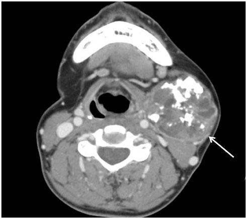
Fig. 1 CT scan of the neck of a 46-year-old female with a history of left submandibular pleomorphic adenoma 9 years prior. Axial post-contrast image reveals a 4.1 × 5.1 cm well-defined non-homogenous enhancing mass in the left submandibular gland with internal calcifications (arrow).
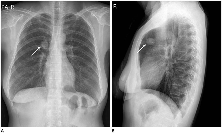
Fig. 2 Chest radiograph of a 46-year-old female with metastasizing pleomorphic adenoma. A, B. Chest radiograph of posterior-anterior projection (A) and right lateral projection (B) show a 2.4 cm, oval, well-defined solitary pulmonary nodule in the right upper lobe (arrows).
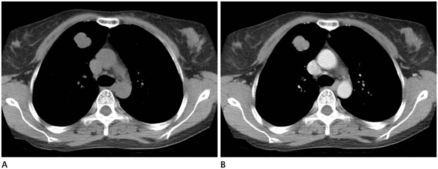
Fig. 3 CT images of metastasizing plemorphic adenoma. A, B. Axial pre-contrast image (A) and post-contrast image (B) show a 2.2 × 2.0 × 2.2 cm well-defined nodule with lobulated margin and relatively homogenous enhancement (12 to 44 Hounsfield units) in the anterior segment of the right upper lobe without cavitation, necrosis, or calcification. CT = computed tomography

Fig. 4 MR images of metastasizing plemorphic adenoma. A-C. Axial T1-weighted (A), T2-weighted (B), and post-contrast enhanced T1-weighted image (C) of the lung show a solitary pulmonary nodule in the anterior segment of the right upper lobe with intermediate to high signal intensity on T1-weighted images, heterogenous high signal intensity on T2-weighted images, and with strong enhancement on gadolinium-enhanced T1-weighted images. MR = magnetic resonance
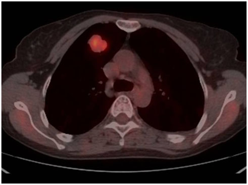
Fig. 5 FDG PET-CT image of metastasizing pleomorphic adenoma. Axial fusion image shows increased FDG uptake (maximum standardized uptake value of 4.98) in the right upper lobe. FDG = fluorodeoxyglucose, PET-CT = positron emission tomography-computed tomography
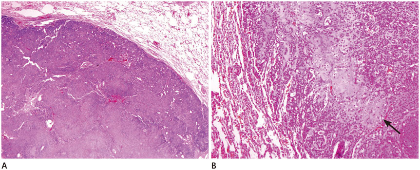
Fig. 6 Microscopic findings of pathologically confirmed metastasizing pleomorphic adenoma. A, B. Low power micrograph (A) of the metastasizing pleomorphic adenoma shows an intrapulmonary well-demarcated cellular nodule (hematoxylin and eosin, × 20). High power micrograph (B) of another field of the metastasizing pleomorphic adenoma shows proliferation of epithelial and myoepithelial cells immersed in a chondromyxoid stroma (arrow) (hematoxylin and eosin, × 100).
Reference
-
1. Rodríguez-Fernández J, Mateos-Micas M, Martínez-Tello FJ, Berjón J, Montalvo JJ, Forteza-González G, et al. Metastatic benign pleomorphic adenoma. Report of a case and review of the literature. Med Oral Patol Oral Cir Bucal. 2008; 13:E193–EE19.2. Sit KY, Chui WH, Wang E, Chiu SW. Multiple pulmonary metastases from benign pleomorphic adenoma. Asian Cardiovasc Thorac Ann. 2008; 16:62–64.3. Zhang Y, Gomez-Fernandez CR, Jorda M, Ganjei-Azar P. Fine-needle aspiration (FNA) and pleural fluid cytology diagnosis of benign metastasizing pleomorphic adenoma of the parotid gland in the lung: a case report and review of literature. Diagn Cytopathol. 2009; 37:828–831.4. Raja V, China C, Masaki KH, Nakano G. Unusual presentations of uncommon tumors: case 1. Benign metastasizing pleomorphic adenoma. J Clin Oncol. 2002; 20:2400–2240.5. Knight J, Ratnasingham K. Metastasising pleomorphic adenoma: systematic review. Int J Surg. 2015; 19:137–145.6. Santaliz-Ruiz LE, Morales G, Santini H, Sánchez-Santiago M, Arroyo A. Metastasizing pleomorphic adenoma: a fascinating enigma. Case Rep Med. 2012; 2012:148103.7. Nouraei SA, Ferguson MS, Clarke PM, Sandison A, Sandhu GS, Michaels L, et al. Metastasizing pleomorphic salivary adenoma. Arch Otolaryngol Head Neck Surg. 2006; 132:788–793.8. Youngs GR, Scheuer PJ. Histologically benign mixed parotid tumour with hepatic metastasis. J Pathol. 1973; 109:171–172.9. Landolt U, Zöbeli L, Pedio G. Pleomorphic adenoma of the salivary glands metastatic to the lung: diagnosis by fine needle aspiration cytology. Acta Cytol. 1990; 34:101–102.10. Kinoshita T, Ishii K, Naganuma H, Okitsu T. MR imaging findings of parotid tumors with pathologic diagnostic clues: a pictorial essay. Clin Imaging. 2004; 28:93–101.
- Full Text Links
-
- Actions
-
Cited
- CITED
-
- Close
- Share
-
- Similar articles
-
- Metastasizing Pleomorphic Adenoma: Report of a Case
- A Case of Aggressive Local Recurrence of Metastasizing Pleomorphic Adenoma with Multiple Lung Metastases
- High-Grade Mucoepidermoid Carcinoma Ex Metastasizing Pleomorphic Adenomas in the Parotid Gland and Parapharyngeal Space: a Case Report and Literature Review
- Pulmonary Pleomorphic Adenoma: Report of a Rare Case
- A Case of Metastasizing Pleomorphic Adenoma Recurred as Cervical Lymph Node Metastasis after Parotidectomy
