Multiple Solitary Plasmacytomas Arising in the Posterior Mediastinum and Sternum: A Case Report
- Affiliations
-
- 1Department of Radiology, Sanggye Paik Hospital, Inje University College of Medicine, Seoul, Korea. s2621@paik.ac.kr
- 2Department of Emergency Medicine, Sanggye Paik Hospital, Inje University College of Medicine, Seoul, Korea.
- KMID: 2155281
- DOI: http://doi.org/10.3348/jksr.2016.74.3.199
Abstract
- Multiple solitary plasmacytoma is very rare disease, which occurs in multiple sites of soft tissue, bone or both without multiple myeloma. Almost 80-90% of extramedullary plasmacytomas develop in the head and neck area. Involvement of the mediastinum is extremely rare. A few cases of mediastinal plasmacytoma have been reported, and to the best of our knowledge, there is only one previous report of multiple plasmacytomas involving the posterior mediastinum. Herein, we describe an unusual case of multiple solitary plasmacytomas in a 67-year-old male involving the posterior mediastinum and sternum without evidence of bone marrow involvement.
MeSH Terms
Figure
-
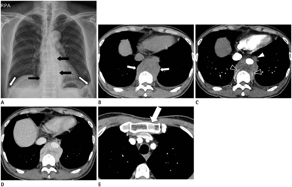
Fig. 1 Plain radiograph and chest CT scan in 67-year-old man with multiple solitary plasmacytomas. Plain radiograph (A) shows thickening of the bilateral paravertebral stripe (black arrows) and bilateral pleural effusion (white arrows). Pre-contrast (B) and post-contrast chest CT (C) images show a heterogeneously enhanced soft tissue mass (white arrows) in the posterior mediastinum at T10 level. Mass encases the abdominal aorta and intercostal arteries (black arrowheads) without definite evidence of invasion. Esophagus (white arrowhead) is replaced anteriorly. Delayed phase of abdominal CT (D) image shows gradual and heterogenous enhancement. Post-contrast CT (E) image shows a round shape osteolytic lesion with sclerotic rim (arrow) in the manubrium of the sternum.
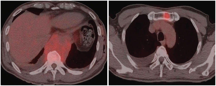
Fig. 2 PET-CT scan in 67-year-old man with multiple solitary plasmacytomas. PET-CT scan shows 18F-FDG hot uptake in the posterior mediastinum (maxSUV 4.6), T10 vertebral body (maxSUV 3.9) and sternal mass (maxSUV 4.4). It also shows hot uptake in the T8 (maxSUV 5.7) and L3 (maxSUV 3.8) vertebrae (not shown). FDG = fluorodeoxyglucose, maxSUV = maximum standardized uptake value, PET-CT = positron emission tomography-computed tomography
Reference
-
1. Ooi GC, Chim JC, Au WY, Khong PL. Radiologic manifestations of primary solitary extramedullary and multiple solitary plasmacytomas. AJR Am J Roentgenol. 2006; 186:821–827.2. Masood A, Hudhud KH, Hegazi A, Syed G. Mediastinal plasmacytoma with multiple myeloma presenting as a diagnostic dilemma. Cases J. 2008; 1:116.3. Lee SY, Kim JH, Shin JS, Shin C, In KH, Kang KH, et al. A case of extramedullary plasmacytoma arising from the posterior mediastinum. Korean J Intern Med. 2005; 20:173–176.4. Hussain A, Singh M, Singh K, Bagga H. Multiple extramedullary plasmacytoma with lytic bony lesions: a rare case report. Case Rep Med. 2013; 2013:291359.5. Ahnach M, Marouan S, Rachid M, Madani A, Quessar A, Benchekroun S, et al. Extramedullary plasmocytoma relapsing at differents sites: an unusual presentation. Pan Afr Med J. 2013; 14:34.6. International Myeloma. Criteria for the classification of monoclonal gammopathies, multiple myeloma and related disorders: a report of the International Myeloma Working Group. Br J Haematol. 2003; 121:749–757.7. Luh SP, Lai YS, Tsai CH, Tsao TC. Extramedullary plasmacytoma (EMP): report of a case manifested as a mediastinal mass and multiple pulmonary nodules and review of literature. World J Surg Oncol. 2007; 5:123.8. Alexiou C, Kau RJ, Dietzfelbinger H, Kremer M, Spiess JC, Schratzenstaller B, et al. Extramedullary plasmacytoma: tumor occurrence and therapeutic concepts. Cancer. 1999; 85:2305–2314.
- Full Text Links
-
- Actions
-
Cited
- CITED
-
- Close
- Share
-
- Similar articles
-
- A Case of Extramedullary Plasmacytomas of Posterior Mediastinum, and Gingiva associated with Fulminant Hepatic Failure, which Developed in the Course of Multiple Myeloma
- Solitary Plasmacytoma of the Sternum
- Multiple Solitary Plasmacytomas Presenting with Painful Erythematous Swelling of the Upper Eyelid
- A Case of Extramedullary Plasmacytoma Arising from the Posterior Mediastinum
- Cutaneous Bronchogenic Cyst Over the Sternum: A Case Report
