A Case Report of the Angiosarcoma Involving Epicranial Muscle and Fascia : Is the Occipitofrontalis Muscle Composed of Two Different Muscles?
- Affiliations
-
- 1Department of Radiology, Catholic University of Daegu School of Medicine, Daegu, Korea.
- 2Department of Radiology, Kyungpook National University Hospital, Daegu, Korea. leehuijoong@knu.ac.kr
- KMID: 2152924
- DOI: http://doi.org/10.3340/jkns.2016.59.1.78
Abstract
- The occipitofrontalis muscle is generally regarded as one muscle composed of two muscle bellies joined through the galea aponeurotica. However, two muscle bellies have different embryological origin, anatomical function and innervations. We report a case of angiosarcoma of the scalp in a 63-year-old man whose MR showed that the superficial fascia overlying the occipital belly becomes the temporoparietal fascia and ends at the superior end of the frontal belly. Beneath the superficial fascia, the occipital belly of the occipitofrontalis muscle becomes the galea aponeurotica and inserts into the underside of the frontal belly. The presented case report supported the concept of which the occipitofrontalis muscle appears to be composed of two anatomically different muscles.
Keyword
MeSH Terms
Figure
-
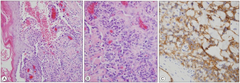
Fig. 1 63-year-old man with angiosarcoma of the scalp. Low power (A, ×100) and high power (B, ×400). Hematoxylin-eosin stain shows pleomorphic endothelial cells, and immunohistochemical staining (C, ×400) shows positive for CD31 antibody staining.
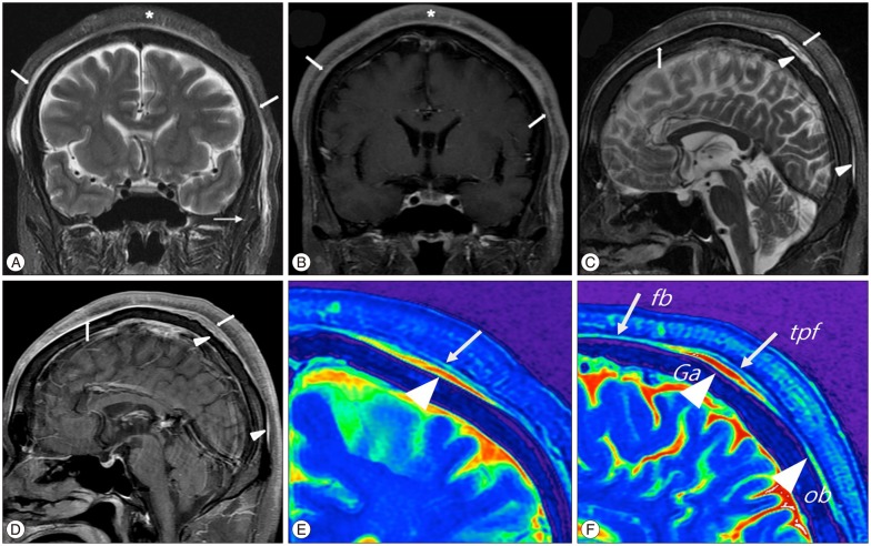
Fig. 2 Coronal view of T2 (A) and contrast enhanced T1 (B) weighted MR image shows a diffuse thickening of the scalp, invading subcutaneous fat with reticular patterns of high signal intensity (asterisk) due to angiosarcoma. Superficial fascia (arrows) shows diffuse thickening and enhancement, covering normal signal intensity of temporalis muscles (small arrow). Sagittal view of T2 (C) and contrast enhanced T1 (D) weighted image show tumor involvement of superficial fascia (arrows), overlying the occipital belly and galea aponeurotica (arrowheads). Color coding image according to signal intensity of coronal (E) and sagittal (F) views of T2 weighted MR image demonstrate the occipital belly of the occipitofrontalis muscle becomes the red colored galea aponeurotica (arrow head) and inserts into the underside of the green colored superficial musculoaponeurotic system (arrows), composed of the frontal belly of occipitofrontalis and temporoparietal fascia. Ga : galea aponeurotica, fb : frontal belly, ob : occipital belly, tpf : temporoparietal fascia.

Fig. 3 PET-CT show diffuse uptake in the scalp (arrowheads) but invisible increased uptake on epicranial muscle (A). Faint uptake is visible in lymph nodes (arrows) at the level VA (B). On the 6 months follow-up PET-CT, hot uptake were visible in the level VA lymph nodes (arrow) which showed faint uptake initial study (C), and multiple lymph node metastases of both neck (arrows) are seen (D).
Reference
-
1. Abul-Hassan HS, von Drasek Ascher G, Acland RD. Surgical anatomy and blood supply of the fascial layers of the temporal region. Plast Reconstr Surg. 1986; 77:17–28. PMID: 3941846.2. Aust MR, Olsen KD, Lewis JE, Nascimento AG, Meland NB, Foote RL, et al. Angiosarcomas of the head and neck : clinical and pathologic characteristics. Ann Otol Rhinol Laryngol. 1997; 106:943–951. PMID: 9373085.
Article3. Bastiaannet E, Groen H, Jager PL, Cobben DC, van der Graaf WT, Vaalburg W, et al. The value of FDG-PET in the detection, grading and response to therapy of soft tissue and bone sarcomas; a systematic review and meta-analysis. Cancer Treat Rev. 2004; 30:83–101. PMID: 14766127.
Article4. Bérzin F. Occipitofrontalis muscle : functional analysis revealed by electromyography. Electromyogr Clin Neurophysiol. 1989; 29:355–358. PMID: 2689156.5. Figueiredo MT, Marques LA, Campos-Filho N. Soft-tissue sarcomas of the head and neck in adults and children : experience at a single institution with a review of literature. Int J Cancer. 1988; 41:198–200. PMID: 3276633.
Article6. Futamura R. Über die entwickelung der fasialismuskulatur des Menschen. Anat Hefte. 1906; 30:436–516.7. Gola R. [The forehead cutaneo-musculo-aponeurotic unit and aging of the forehead. Anatomo-physiological considerations and surgical implications]. Ann Chir Plast Esthet. 1999; 44:89–102. PMID: 10188299.8. Hayman LA, Shukla V, Ly C, Taber KH. Clinical and imaging anatomy of the scalp. J Comput Assist Tomogr. 2003; 27:454–459. PMID: 12826816.
Article9. Isoda H, Imai M, Inagawa S, Miura K, Sakahara H. Magnetic resonance imaging findings of angiosarcoma of the scalp. J Comput Assist Tomogr. 2005; 29:858–862. PMID: 16272865.
Article10. Kushima H, Matsuo K, Yuzuriha S, Kitazawa T, Moriizumi T. The occipitofrontalis muscle is composed of two physiologically and anatomically different muscles separately affecting the positions of the eyebrow and hairline. Br J Plast Surg. 2005; 58:681–687. PMID: 15927153.
Article11. Mark RJ, Tran LM, Sercarz J, Fu YS, Calcaterra TC, Juillard GF. Angiosarcoma of the head and neck. The UCLA experience 1955 through 1990. Arch Otolaryngol Head Neck Surg. 1993; 119:973–978. PMID: 8357598.
Article12. Pawlik TM, Paulino AF, McGinn CJ, Baker LH, Cohen DS, Morris JS, et al. Cutaneous angiosarcoma of the scalp : a multidisciplinary approach. Cancer. 2003; 98:1716–1726. PMID: 14534889.13. Rosai J, Sumner HW, Kostianovsky M, Perez-Mesa C. Angiosarcoma of the skin. A clinicopathologic and fine structural study. Hum Pathol. 1976; 7:83–109. PMID: 942663.14. Standring S. Gray's anatomy. London: Churchill Livingstone;2004.
- Full Text Links
-
- Actions
-
Cited
- CITED
-
- Close
- Share
-
- Similar articles
-
- Synovial Sarcoma in the Calf Muscle Fascia: A Case Report
- Anterior Tibial Muscle Hernia Treated with Local Periosteal Rotational Flap: A Case Report
- Effect of Kinesio Tape for Fascia on Trunk Muscle Activity during Plank
- Usefulness of Ultrasonographic Examination of Diagnosis of Muscle Hernia
- A Modified Under-Vastus Approach for Knee Arthroplasty with Anatomical Repair of Soft Tissue
