A comparison of the precision of three-dimensional images acquired by 2 digital intraoral scanners: effects of tooth irregularity and scanning direction
- Affiliations
-
- 1Department of Orthodontics, Graduate School of Clinical Dentistry, Ewha Womans University, Seoul, Korea. minjikim@ewha.ac.kr
- 2Department of Prosthodontics and Dental Research Institute, Seoul National University Gwanak Dental Hospital, Seoul, Korea.
- 3Department of Pediatric Dentistry, Graduate School of Clinical Dentistry, Ewha Womans University, Seoul, Korea.
- KMID: 2152892
- DOI: http://doi.org/10.4041/kjod.2016.46.1.3
Abstract
OBJECTIVE
The purpose of this study was to compare the precision of three-dimensional (3D) images acquired using iTero(R) (Align Technology Inc., San Jose, CA, USA) and Trios(R) (3Shape Dental Systems, Copenhagen, Denmark) digital intraoral scanners, and to evaluate the effects of the severity of tooth irregularities and scanning sequence on precision.
METHODS
Dental arch models were fabricated with differing degrees of tooth irregularity and divided into 2 groups based on scanning sequence. To assess their precision, images were superimposed and an optimized superimposition algorithm was employed to measure any 3D deviation. The t-test, paired t-test, and one-way ANOVA were performed (p < 0.05) for statistical analysis.
RESULTS
The iTero(R) and Trios(R) systems showed no statistically significant difference in precision among models with differing degrees of tooth irregularity. However, there were statistically significant differences in the precision of the 2 scanners when the starting points of scanning were different. The iTero(R) scanner (mean deviation, 29.84 +/- 12.08 microm) proved to be less precise than the Trios(R) scanner (22.17 +/- 4.47 microm).
CONCLUSIONS
The precision of 3D images differed according to the degree of tooth irregularity, scanning sequence, and scanner type. However, from a clinical standpoint, both scanners were highly accurate regardless of the degree of tooth irregularity.
Keyword
MeSH Terms
Figure
-
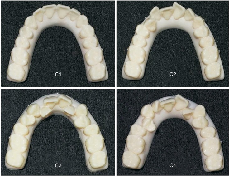
Figure 1 Dental arch models according to the severity of tooth irregularity expressed as arch length discrepancy (ALD). C1, Ideal arch dentition (ALD, 0 mm); C2, mildly crowded dentition (ALD, 3 mm); C3, moderately crowded dentition (ALD, 7 mm); C4, severely crowded dentition (ALD, 10 mm). Fiducials with a diameter of 1.5 mm were attached at 5 locations on the model base located 4 mm below the dentogingival junction. Two posterior fiducials were located below the mesiobuccal cusp of the first molars and 1 anterior ball marker was located below the contact point between the central incisors. Two canine fiducials were located directly below the cusp tips of the right and left canine teeth.
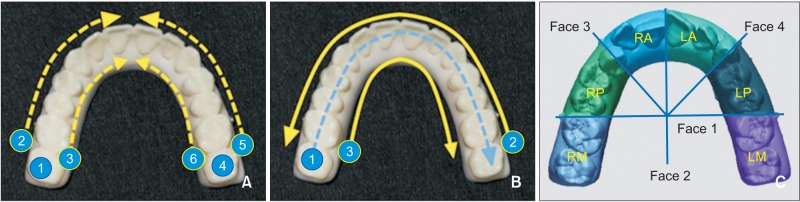
Figure 2 Scanning sequences and reference faces for model division and the model sections. A, The Group Right sequence on the iTero®. The right molars were scanned first in the order of the occlusal side ①, buccal side ②, and palatal side ③; subsequently, the left molars were scanned in the same fashion in the order of ④, ⑤, and ⑥. Both scanned images were merged and connected at the anterior area. The scanning sequence for Group Left first scanned the left molars in reverse. B, The sequence for Group Right on the Trios®. Scanning started from the occlusal side of the right molars and proceeded toward the left occlusal side ①, continued along the buccal side in the reverse direction ②, and lastly, from the palatal side of the right molars, proceeding toward the palatal side of the left molars ③. The scanning sequence for Group Left first scanned the left side in reverse. C, The reference faces for model division and the model sections. Face 1 is a plane passing through the posterior fiducials on both sides, Face 2 is a plane passing through the anterior ball marker and perpendicular to Face 1, and Faces 3 and 4 are planes passing the fiducials in the canine region and the intersection point of Faces 1 and 2.The scanned images were divided into 6 sections: RM, right molar; RP, right premolar; RA, right anterior; LM, left molar; LP, left premolar; LA, left anterior.
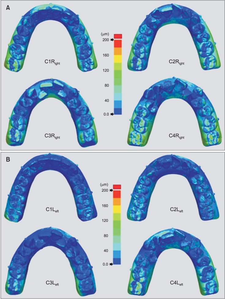
Figure 3 A color presentation of the deviations between surfaces from the iTero® scanner. The color map was set to range from 0 µm to +200 µm. For the models in upper box A (C1Right, C2Right, C3Right, and C4Right), scanning started in the right molar region and then continued on the left side; in lower box B (C1Left, C2Left, C3Left, and C4Left), scanning started in the left molar region and then continued on the right side.C1, Ideal arch dentition; C2, mildly crowded dentition; C3, moderately crowded dentition; C4, severely crowded dentition.
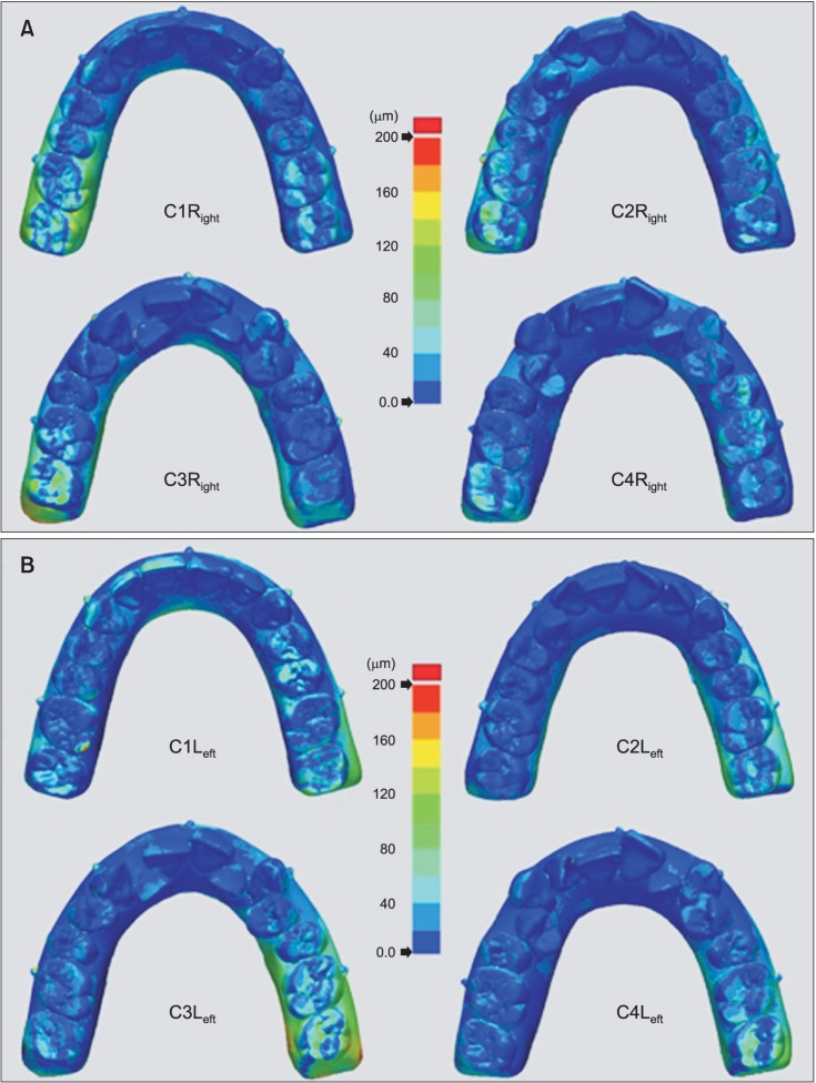
Figure 4 A color presentation of the deviations between surfaces from the Trios® scanner. The color map was set to range from 0 µm to +200 µm. For the models in upper box A (C1Right, C2Right, C3Right, and C4Right), scanning started on the occlusal side of the right molar and continued toward the left side; in lower box B (C1Left, C2Left, C3Left, and C4Left), scanning started on the occlusal side of the left molar and continued toward the right side.C1, Ideal arch dentition; C2, mildly crowded dentition; C3, moderately crowded dentition; C4, severely crowded dentition.
Cited by 3 articles
-
Clinical application of an intraoral scanner for serial evaluation of orthodontic tooth movement: A preliminary study
Dalsun Yun, Dong-Soon Choi, Insan Jang, Bong-Kuen Cha
Korean J Orthod. 2018;48(4):262-267. doi: 10.4041/kjod.2018.48.4.262.Comparison of patient satisfaction with digital and conventional impression for prosthodontic treatment
Hyung-In Yoon, Su-Min Lee, Eun-Jin Park
J Korean Acad Prosthodont. 2016;54(4):379-386. doi: 10.4047/jkap.2016.54.4.379.Evaluation of accuracy of 3-dimensional printed dental models in reproducing intermaxillary relational measurements: Based on inter-operator differences
Won-joon Choi, Su-jung Lee, Cheol-Hyun Moon
Korean J Orthod. 2022;52(1):20-28. doi: 10.4041/kjod.2022.52.1.20.
Reference
-
1. Mah J, Hatcher D. Current status and future needs in craniofacial imaging. Orthod Craniofac Res. 2003; 6(Suppl 1):10–16. PMID: 14606529.
Article2. White AJ, Fallis DW, Vandewalle KS. Analysis of intra-arch and interarch measurements from digital models with 2 impression materials and a modeling process based on cone-beam computed tomography. Am J Orthod Dentofacial Orthop. 2010; 137:456.e1–456.e9. PMID: 20362900.
Article3. Chandran DT, Jagger DC, Jagger RG, Barbour ME. Two- and three-dimensional accuracy of dental impression materials: effects of storage time and moisture contamination. Biomed Mater Eng. 2010; 20:243–249. PMID: 21084736.
Article4. Al Mortadi N, Eggbeer D, Lewis J, Williams RJ. CAD/CAM/AM applications in the manufacture of dental appliances. Am J Orthod Dentofacial Orthop. 2012; 142:727–733. PMID: 23116514.
Article5. Beuer F, Schweiger J, Edelhoff D. Digital dentistry: an overview of recent developments for CAD/CAM generated restorations. Br Dent J. 2008; 204:505–511. PMID: 18469768.
Article6. Cha BK, Lee JY, Jost-Brinkmann PG, Yoshida N. Analysis of tooth movement in extraction cases using three-dimensional reverse engineering technology. Eur J Orthod. 2007; 29:325–331. PMID: 17513876.
Article7. Hajeer MY, Millett DT, Ayoub AF, Siebert JP. Applications of 3D imaging in orthodontics: part II. J Orthod. 2004; 31:154–162. PMID: 15210932.8. Patel N. Integrating three-dimensional digital technologies for comprehensive implant dentistry. J Am Dent Assoc. 2010; 141(Suppl 2):20S–24S. PMID: 20516111.
Article9. Zhang XJ, He L, Guo HM, Tian J, Bai YX, Li S. Integrated three-dimensional digital assessment of accuracy of anterior tooth movement using clear aligners. Korean J Orthod. 2015; 45:275–281. PMID: 26629473.
Article10. Watanabe-Kanno GA, Abrão J, Miasiro Junior H, Sánchez-Ayala A, Lagravère MO. Reproducibility, reliability and validity of measurements obtained from Cecile3 digital models. Braz Oral Res. 2009; 23:288–295. PMID: 19893964.
Article11. Yourtee D, Emery J, Smith RE, Hodgson B. Stereolithographic models of biopolymers. J Mol Graph Model. 2000; 18:26–28. 59–60. PMID: 10935203.
Article12. Normung DDIf. Accuracy (trueness and precision) of measurement methods and results - Part 1: General principles and definitions (ISO 5725-1:1994). Berlin: Beuth Verlag GmbH;1997.13. Ender A, Mehl A. Accuracy of complete-arch dental impressions: a new method of measuring trueness and precision. J Prosthet Dent. 2013; 109:121–128. PMID: 23395338.
Article14. Flügge TV, Schlager S, Nelson K, Nahles S, Metzger MC. Precision of intraoral digital dental impressions with iTero and extraoral digitization with the iTero and a model scanner. Am J Orthod Dentofacial Orthop. 2013; 144:471–478. PMID: 23992820.
Article15. Wiranto MG, Engelbrecht WP, Tutein Nolthenius HE, van der Meer WJ, Ren Y. Validity, reliability, and reproducibility of linear measurements on digital models obtained from intraoral and cone-beam computed tomography scans of alginate impressions. Am J Orthod Dentofacial Orthop. 2013; 143:140–147. PMID: 23273370.
Article16. Naidu D, Freer TJ. Validity, reliability, and reproducibility of the iOC intraoral scanner: a comparison of tooth widths and Bolton ratios. Am J Orthod Dentofacial Orthop. 2013; 144:304–310. PMID: 23910212.
Article17. Patzelt SB, Emmanouilidi A, Stampf S, Strub JR, Att W. Accuracy of full-arch scans using intraoral scanners. Clin Oral Investig. 2014; 18:1687–1694.
Article18. Persson AS, Odén A, Andersson M, Sandborgh-Englund G. Digitization of simulated clinical dental impressions: virtual three-dimensional analysis of exactness. Dent Mater. 2009; 25:929–936. PMID: 19264353.
Article19. Mehl A, Ender A, Mörmann W, Attin T. Accuracy testing of a new intraoral 3D camera. Int J Comput Dent. 2009; 12:11–28. PMID: 19213357.20. Logozzo S, Zanetti EM, Franceschini G, Kilpelä A, Mäkynen A. Recent advances in dental optics-Part I: 3D intraoral scanners for restorative dentistry. Opt Laser Eng. 2014; 54:203–221.21. Kusnoto B, Evans CA. Reliability of a 3D surface laser scanner for orthodontic applications. Am J Orthod Dentofacial Orthop. 2002; 122:342–348. PMID: 12411877.
Article22. Ender A, Mehl A. Full arch scans: conventional versus digital impressions--an in-vitro study. Int J Comput Dent. 2011; 14:11–21. PMID: 21657122.23. Ender A, Mehl A. Influence of scanning strategies on the accuracy of digital intraoral scanning systems. Int J Comput Dent. 2013; 16:11–21. PMID: 23641661.
- Full Text Links
-
- Actions
-
Cited
- CITED
-
- Close
- Share
-
- Similar articles
-
- Comparative analysis on reproducibility among 5 intraoral scanners: sectional analysis according to restoration type and preparation outline form
- Full-arch accuracy of five intraoral scanners:In vivo analysis of trueness and precision
- Effect of scanning strategies on the accuracy of digital intraoral scanners: a meta-analysis of in vitro studies
- Comparison of intraoral scanning and conventional impression techniques using 3-dimensional superimposition
- Quantitative and qualitative evaluation on the accuracy of three intraoral scanners for human identification in forensic odontology
