In silico analysis of an envelope domain III-based multivalent fusion protein as a potential dengue vaccine candidate
- Affiliations
-
- 1Department of Molecular and Cellular Sciences, Faculty of Advanced Sciences and Technology, Pharmaceutical Sciences Branch, Islamic Azad University (IAUPS), Tehran, Iran.
- 2Department of Molecular Genetics, Faculty of Biological Sciences, Tarbiat Modares University, Tehran, Iran. sadeghma@modares.ac.ir
- 3Blood Transfusion Research Center, High Institute for Research and Education in Transfusion Medicine, Tehran, Iran.
- KMID: 2152696
- DOI: http://doi.org/10.7774/cevr.2016.5.1.41
Abstract
- PURPOSE
Dengue virus infection is now a global problem. Currently, there is no licensed vaccine or proven antiviral treatment against this virus. All four serotypes (1-4) of dengue virus can infect human. An effective dengue vaccine should be tetravalent to induce protective immune responses against all four serotypes. Most of dengue vaccine candidates are monovalent, or in the form of physically mixed multivalent formulations. Recently envelope protein domain III of virus is considered as a vaccine candidate, which plays critical roles in the most important viral activities. Development of a tetravalent protein subunit vaccine is very important for equal induction of immune system and prevention of unbalanced immunity. Here, we have presented and used a rational approach to design a tetravalent dengue vaccine candidate.
MATERIALS AND METHODS
We designed a multi domain antigen by fusing four consensus domain III sequences together with appropriate hydrophobic linkers and used several types of bioinformatics software and neural networks to predict structural and immunological properties of the designed tetravalent antigen.
RESULTS
We designed a tetravalent protein (EDIIIF) based on domain III of dengue virus envelope protein. According to the results of the bioinformatics analysis, the constructed models for EDIIIF protein were structurally stable and potentially immunogenic.
CONCLUSION
The designed tetravalent protein can be considered as a potential dengue vaccine candidate. The presented approach can be used for rational design and in silico evaluation of chimeric dengue vaccine candidates.
Keyword
MeSH Terms
Figure
-

Fig. 1 A schematic representation for genomic organization of the dengue virus. The long genomic RNA contains an open reading frame and flanked by 5' and 3' non-coding regions (NCRs), which are shown at the either end. The coding regions of 10 viral proteins are shown by green and brown boxes.
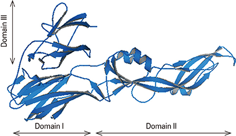
Fig. 2 Three dimensional structure of monomeric form of dengue virus E protein [7]. The potent neutralizing epitopes are located in domain III.
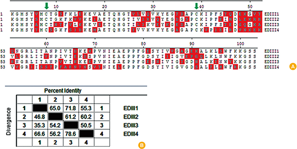
Fig. 3 Comparison of amino acid sequences of the four consensus EDIIIs. (A) Sequence alignment of EDIIIs; different amino acid residues between serotypes are indicated in red blocks. The most conserves cysteine residues are showed by green arrows in the positions of 8 and 39. (B) Percentage identity and divergence among EDIIIs of four serotypes.

Fig. 4 Schematic representation of the EDIIIF construct.
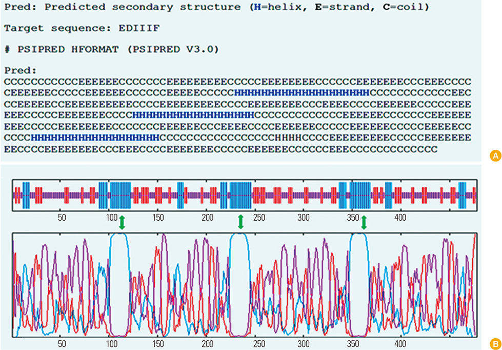
Fig. 5 Prediction of EDIIIF protein secondary structure by PSIpred (A) and GOR4 (B) methods. (A) Formation of α-helix structures in linker segments are showed by H (blue). As described previously for native structure of envelope domain III, each domain contains several β-sheets and coils, which are depicted by the letters of E and C, respectively. (B) The predicted corresponding positions of α-helix structures depicted by arrows in three regions.
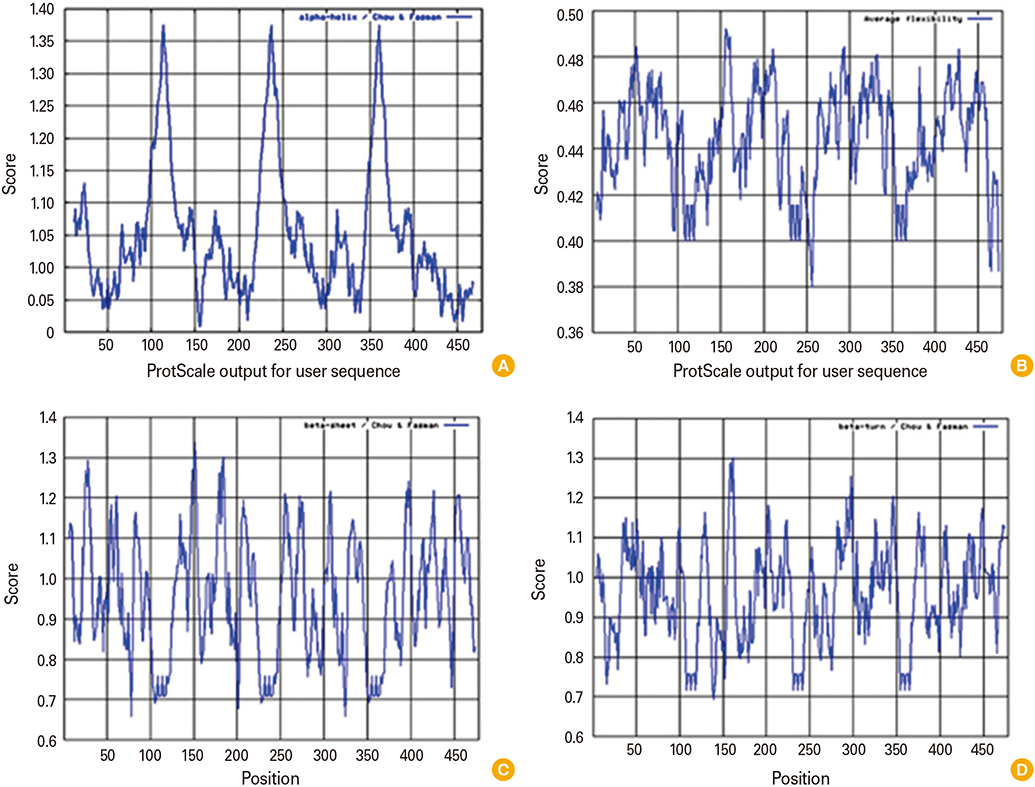
Fig. 6 Predicted properties of EDIIIF protein in secondary structure by using Chou and Fasman method in ProtScale server. The scores for formation of α-helix structures (A), the average flexibility (B), beta-sheets (C), and beta turns (D) throughout the EDIIIF sequence are showed.
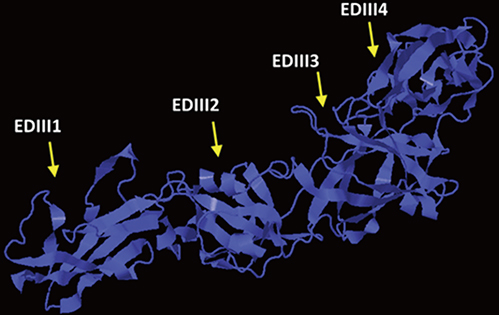
Fig. 7 Homology modeling was used to predict the tertiary structure of the EDIIIF protein. All Four separated EDIII domains (EDIII1, EDIII2, EDIII3, and EDIII4) were presented by arrows. The results were viewed by PyMOL software.

Fig. 8 Evaluation of model stability by using the Ramachandran plot. According to the plot statistics, more than 82% of amino acid residues are in the most favored regions (A, B, L), and 13% are in additional allowed regions (a, b, l, p); whereas only 2.7% are in generously allowed (-a, -b, -l, -p) and 1.7% are in disallowed regions. Accordingly, the constructed model has a good quality.

Fig. 9 Prediction of relative solvent accessibility by using Scratch server.
Reference
-
1. Kuno G, Chang GJ, Tsuchiya KR, Karabatsos N, Cropp CB. Phylogeny of the genus Flavivirus. J Virol. 1998; 72:73–83.2. Halstead SB. Dengue. Lancet. 2007; 370:1644–1652.
Article3. Rothman AL. Dengue: defining protective versus pathologic immunity. J Clin Invest. 2004; 113:946–951.
Article4. Fields BN, Knipe DM, Howley PM. Fields' virology. 5th ed. Philadelphia: Wolters Kluwer Health/Lippincott Williams & Wilkins;2007.5. Crill WD, Chang GJ. Localization and characterization of flavivirus envelope glycoprotein cross-reactive epitopes. J Virol. 2004; 78:13975–13986.
Article6. Roehrig JT, Bolin RA, Kelly RG. Monoclonal antibody mapping of the envelope glycoprotein of the dengue 2 virus, Jamaica. Virology. 1998; 246:317–328.
Article7. Modis Y, Ogata S, Clements D, Harrison SC. Structure of the dengue virus envelope protein after membrane fusion. Nature. 2004; 427:313–319.
Article8. Crill WD, Roehrig JT. Monoclonal antibodies that bind to domain III of dengue virus E glycoprotein are the most efficient blockers of virus adsorption to Vero cells. J Virol. 2001; 75:7769–7773.
Article9. Chavez JH, Silva JR, Amarilla AA, Moraes Figueiredo LT. Domain III peptides from flavivirus envelope protein are useful antigens for serologic diagnosis and targets for immunization. Biologicals. 2010; 38:613–618.
Article10. Swaminathan S, Batra G, Khanna N. Dengue vaccines: state of the art. Expert Opin Ther Pat. 2010; 20:819–835.
Article11. Puigbo P, Guzman E, Romeu A, Garcia-Vallve S. OPTIMIZER: a web server for optimizing the codon usage of DNA sequences. Nucleic Acids Res. 2007; 35:W126–W131.
Article12. Vincze T, Posfai J, Roberts RJ. NEBcutter: a program to cleave DNA with restriction enzymes. Nucleic Acids Res. 2003; 31:3688–3691.
Article13. Zuker M. Mfold web server for nucleic acid folding and hybridization prediction. Nucleic Acids Res. 2003; 31:3406–3415.
Article14. Garnier J, Gibrat JF, Robson B. GOR method for predicting protein secondary structure from amino acid sequence. Methods Enzymol. 1996; 266:540–553.15. Arai R, Ueda H, Kitayama A, Kamiya N, Nagamune T. Design of the linkers which effectively separate domains of a bifunctional fusion protein. Protein Eng. 2001; 14:529–532.
Article16. Amani J, Mousavi SL, Rafati S, Salmanian AH. In silico analysis of chimeric espA, eae and tir fragments of Escherichia coli O157:H7 for oral immunogenic applications. Theor Biol Med Model. 2009; 6:28.
Article17. Koraka P, Martina BE, Osterhaus AD. Bioinformatics in new generation flavivirus vaccines. J Biomed Biotechnol. 2010; 2010:864029.
Article18. Holbrook MR, Shope RE, Barrett AD. Use of recombinant E protein domain III-based enzyme-linked immunosorbent assays for differentiation of tick-borne encephalitis serocomplex flaviviruses from mosquito-borne flaviviruses. J Clin Microbiol. 2004; 42:4101–4110.
Article19. Fahimi H, Allahyari H, Hassan ZM, Sadeghizadeh M. Dengue virus type-3 envelope protein domain III; expression and immunogenicity. Iran J Basic Med Sci. 2014; 17:836–843.20. Burgess-Brown NA, Sharma S, Sobott F, Loenarz C, Oppermann U, Gileadi O. Codon optimization can improve expression of human genes in Escherichia coli: a multi-gene study. Protein Expr Purif. 2008; 59:94–102.
Article21. Tong JC, Tan TW, Ranganathan S. Methods and protocols for prediction of immunogenic epitopes. Brief Bioinform. 2007; 8:96–108.
Article22. Kolaskar AS, Tongaonkar PC. A semi-empirical method for prediction of antigenic determinants on protein antigens. FEBS Lett. 1990; 276:172–174.
Article
- Full Text Links
-
- Actions
-
Cited
- CITED
-
- Close
- Share
-
- Similar articles
-
- Prospects for dengue vaccines for travelers
- A potent multivalent vaccine for modulation of immune system in atherosclerosis: an in silico approach
- Development of a Novel Subunit Vaccine Targeting Fusobacterium nucleatum FomA Porin Based on In Silico Analysis
- Animal models for dengue vaccine development and testing
- Identifying immunodominant multi-epitopes from the envelope glycoprotein of the Lassa mammarenavirus as vaccine candidate for Lassa fever
