Obstet Gynecol Sci.
2016 Jan;59(1):66-70. 10.5468/ogs.2016.59.1.66.
Intra-amniotic thyroxine to treat fetal goiter
- Affiliations
-
- 1Department of Obstetrics and Gynecology, Cheil General Hospital and Women's Healthcare Center, Dankook University College of Medicine, Seoul, Korea. mykimdr@yahoo.com
- 2Department of Obstetrics and Gynecology, Bundang Cheil Women's Healthcare Center, Seongnam, Korea.
- 3Division of Endocrinology and Metabolism, Cheil General Hospital and Women's Healthcare Center, Dankook University College of Medicine, Seoul, Korea.
- KMID: 2152648
- DOI: http://doi.org/10.5468/ogs.2016.59.1.66
Abstract
- A 35-year-old pregnant woman visited our department and had been treated with 100 microg of daily oral levothyroxine for hypothyroidism. An ultrasonography screening was performed at 25 weeks gestation and revealed a fetal goiter and an increased amniotic fluid volume. Fetal hypothyroidism was confirmed by cordocentesis and amniotic hormone levels at 26 weeks gestation. We treated the mother with 200 microg of daily oral levothyroxine to optimize the transplacental transfer. A total of four intra-amniotic injections of levothyroxine were administered, resulting in progressive reduction in the fetal thyroid volume of goiter as measured by 3D ultrasonography and increased amniotic fluid volume. Following birth, neonatal serum thyroid stimulating hormone level was within the normal range, but free T4 was reduced. Based on this case, we suggest that monitoring amniotic fluid thyroid hormone concentration and intra-amniotic levothyroxine injection can be used to reduce the thyroid volume of goiters and to prevent polyhydramnios.
MeSH Terms
Figure
-
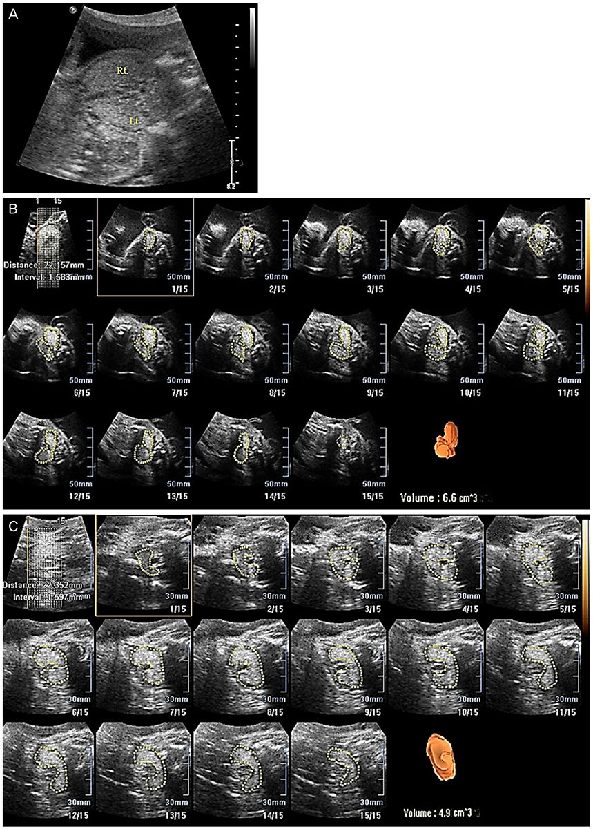
Fig. 1 Measurement of fetal goiter volume change using 2D and 3D ultrasonography. (A) Fetal goiter size at 25 weeks using 2D ultrasonography. Right (Rt) lobe (21×14) and left (Lt) lobe (26×22). (B) Fetal thyroid volume (6.6 cm3) at 29 weeks' gestation using 3D ultrasonography. (C) Fetal thyroid volume (4.9 cm3) at 37 weeks' gestation using 3D ultrasonography.
Reference
-
1. Dussault JH, Coulombe P, Laberge C, Letarte J, Guyda H, Khoury K. Preliminary report on a mass screening program for neonatal hypothyroidism. J Pediatr. 1975; 86:670–674.2. Davidson KM, Richards DS, Schatz DA, Fisher DA. Successful in utero treatment of fetal goiter and hypothyroidism. N Engl J Med. 1991; 324:543–546.3. Perelman AH, Johnson RL, Clemons RD, Finberg HJ, Clewell WH, Trujillo L. Intrauterine diagnosis and treatment of fetal goitrous hypothyroidism. J Clin Endocrinol Metab. 1990; 71:618–621.4. Becks GP, Burrow GN. Thyroid disease and pregnancy. Med Clin North Am. 1991; 75:121–150.5. Bernardes LS, Ruano R, Sapienza AD, Maganha CA, Zugaib M. Nomograms of fetal thyroid measurements estimated by 2-dimensional sonography. J Clin Ultrasound. 2008; 36:193–199.6. Glorieux J, Dussault J, Van Vliet G. Intellectual development at age 12 years of children with congenital hypothyroidism diagnosed by neonatal screening. J Pediatr. 1992; 121:581–584.7. Kooistra L, Laane C, Vulsma T, Schellekens JM, van der Meere JJ, Kalverboer AF. Motor and cognitive development in children with congenital hypothyroidism: a long-term evaluation of the effects of neonatal treatment. J Pediatr. 1994; 124:903–909.8. Ho SS, Metreweli C. Normal fetal thyroid volume. Ultrasound Obstet Gynecol. 1998; 11:118–122.9. Francois A, Hindryckx A, Vandecruys H, Van Schoubroeck D, Vanhole C, Allegaert K, et al. Fetal treatment for early dyshormonogenetic goiter. Prenat Diagn. 2009; 29:543–545.10. Hanono A, Shah B, David R, Buterman I, Roshan D, Shah S, et al. Antenatal treatment of fetal goiter: a therapeutic challenge. J Matern Fetal Neonatal Med. 2009; 22:76–80.11. Corral E, Reascos M, Preiss Y, Rompel SM, Sepulveda W. Treatment of fetal goitrous hypothyroidism: value of direct intramuscular L-thyroxine therapy. Prenat Diagn. 2010; 30:899–901.12. Marin RC, Bello-Munoz JC, Martinez GV, Martinez SA, Moratonas EC, Roura LC. Use of 3-dimensional sonography for prenatal evaluation and follow-up of fetal goitrous hypothyroidism. J Ultrasound Med. 2010; 29:1339–1343.13. Nath CA, Oyelese Y, Yeo L, Chavez M, Kontopoulos EV, Giannina G, et al. Three-dimensional sonography in the evaluation and management of fetal goiter. Ultrasound Obstet Gynecol. 2005; 25:312–314.
- Full Text Links
-
- Actions
-
Cited
- CITED
-
- Close
- Share
-
- Similar articles
-
- Prenatal Diagnosis of Fetal Goiter in a Euthyroid Mother
- A Case of Intrauterine Thyroxine Therapy for Fetal Goitrous Hypothyroidsm
- A Case of Prenatal Diagnosis of Congenital Fetal Goiter in Hyperthyroidism Mother
- A case of congenital goiter with congenital hypothyroidism due to organification defect
- Pendred's Syndrome
