Duodenal Metastasis of Pulmonary Pleomorphic Carcinoma: A Case Report
- Affiliations
-
- 1Department of Radiology, Soonchunhyang University Bucheon Hospital, Bucheon, Korea. jspark@schmc.ac.kr
- 2Purun Radiologic Clinic, Yesan, Korea.
- 3Department of Pathology, Soonchunhyang University Bucheon Hospital, Bucheon, Korea.
- 4Department of Thoracic Surgery, Soonchunhyang University Bucheon Hospital, Bucheon, Korea.
- KMID: 2152600
- DOI: http://doi.org/10.3348/jksr.2016.74.2.91
Abstract
- Pulmonary pleomorphic carcinoma is an uncommon malignant lesion of the lung. A chest radiograph of 53-year-old man who was suffering from a cough revealed a well-defined mass-like opacity with a broad base on the pleura at the apico-posterior segment of the right upper lobe of the lung. The subsequent chest computed tomography (CT) scan demonstrated an inhomogeneous enhancing mass with central low-attenuation in the right upper lobe. A lobectomy was performed and the mass was determined to be a pleomorphic carcinoma with visceral pleura invasion. Forty days after the operation, the patient complained of melena and an abdominal CT revealed an intraluminal and extraluminal protruding mass around the prepyloric antrum and duodenal bulb. The mass was removed by en-block surgery and diagnosed as metastatic pleomorphic carcinoma from the lung. Previous articles reported a median survival time of 3-10 months for pleomorphic carcinoma, but in this case, the patient has continued to survive, 11 years after surgery. Chest and abdominal CTs have revealed no evidence of tumor recurrence or metastasis.
MeSH Terms
Figure
-

Fig. 1 Pleomorphic carcinoma of the lung in a 53-year-old man. A. A chest postero-anterior radiograph reveals a well-defined mass with a maximum diameter of 8.2 cm (arrows) in the right upper lobe. B. A transverse contrast-enhanced CT scan shows a 5.7 cm sized, inhomogeneously enhancing mass lesion with inner low attenuated area (star) in the peripheral lung area of the right upper lobe. The mass abutted the adjacent visceral pleura (arrow). C, D. Photomicrographs of histopathologic specimens (hematoxylin and eosin staining, × 200) show a mixed composition of spindle cell carcinoma (C) and giant cell carcinoma (D).
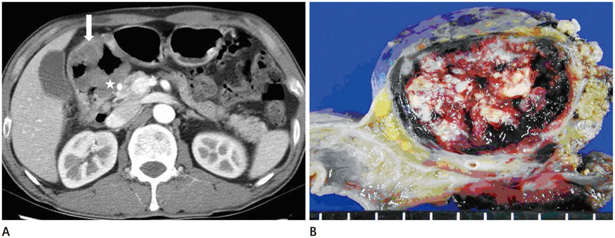
Fig. 2 Duodenal metastasis of pulmonary pleomorphic carcinoma. A. A transverse contrast-enhanced abdominal CT scan shows an intraluminal (arrow) and extraluminal (star) protruding mass around prepyloric antrum and duodenal bulb. B. A photograph of gross specimen of duodenum reveals well-defined serosal mass with extensive hemorrhagic necrosis and mucosal ulceration.
Reference
-
1. Brambilla E, Travis WD, Colby TV, Corrin B, Shimosato Y. The new World Health Organization classification of lung tumours. Eur Respir J. 2001; 18:1059–1068.2. Kim TH, Kim SJ, Ryu YH, Lee HJ, Goo JM, Im JG, et al. Pleomorphic carcinoma of lung: comparison of CT features and pathologic findings. Radiology. 2004; 232:554–559.3. Kim TS, Han J, Lee KS, Jeong YJ, Kwak SH, Byun HS, et al. CT findings of surgically resected pleomorphic carcinoma of the lung in 30 patients. AJR Am J Roentgenol. 2005; 185:120–125.4. Chang YL, Lee YC, Shih JY, Wu CT. Pulmonary pleomorphic (spindle) cell carcinoma: peculiar clinicopathologic manifestations different from ordinary non-small cell carcinoma. Lung Cancer. 2001; 34:91–97.5. Yoo SH, Han J, Kim TJ, Chung DH. Expression of CD99 in pleomorphic carcinomas of the lung. J Korean Med Sci. 2005; 20:50–55.6. Rossi G, Cavazza A, Sturm N, Migaldi M, Facciolongo N, Longo L, et al. Pulmonary carcinomas with pleomorphic, sarcomatoid, or sarcomatous elements: a clinicopathologic and immunohistochemical study of 75 cases. Am J Surg Pathol. 2003; 27:311–324.7. Fishback NF, Travis WD, Moran CA, Guinee DG Jr, McCarthy WF, Koss MN. Pleomorphic (spindle/giant cell) carcinoma of the lung. A clinicopathologic correlation of 78 cases. Cancer. 1994; 73:2936–2945.8. Raveglia F, Mezzetti M, Panigalli T, Furia S, Giuliani L, Conforti S, et al. Personal experience in surgical management of pulmonary pleomorphic carcinoma. Ann Thorac Surg. 2004; 78:1742–1747.9. Segawa M, Kusajima Y, Saito K. [Pleomorphic carcinoma of the lung rapidly developed multiple metastases after surgery]. Kyobu Geka. 2006; 59:387–391.10. Aketa A, Yamada G, Aketa K, Ohnishi T, Takahashi Y, Kudoh K, et al. [Two younger male patients with rapidly progressing pulmonary pleomorphic carcinoma]. Nihon Kokyuki Gakkai Zasshi. 2004; 42:164–169.
- Full Text Links
-
- Actions
-
Cited
- CITED
-
- Close
- Share
-
- Similar articles
-
- Multimodality Imaging of Metastasizing Pleomorphic Adenoma Presenting as a Solitary Pulmonary Nodule without Local Tumor Recurrence: A Case Report
- A CASE OF CARCINOMA EX PLEOMORPHIC ADENOMA OF PALATE
- High-Grade Mucoepidermoid Carcinoma Ex Metastasizing Pleomorphic Adenomas in the Parotid Gland and Parapharyngeal Space: a Case Report and Literature Review
- A Case of Carcinoma Ex Pleomorphic Adenoma in the Maxillary Sinus
- Pulmonary Pleomorphic Adenoma: Report of a Rare Case
