A Case of Hypoglycemic Brain Injuries with Cortical Laminar Necrosis
- Affiliations
-
- 1Department of Neurosurgery, Hangang Sacred Heart Hospital, College of Medicine, Hallym University, Seoul, Korea. neuri71@gmail.com
- 2Division of Cardiology, Department of Internal Medicine, College of Medicine, University of Ulsan, Seoul, Korea.
- 3Division of Endocrinology and Metabolism, Department of Internal Medicine, College of Medicine, Hallym University, Seoul, Korea.
- KMID: 2150881
- DOI: http://doi.org/10.3346/jkms.2010.25.6.961
Abstract
- We report a case of 68-yr-old male who died from brain injuries following an episode of prolonged hypoglycemia. While exploring controversies surrounding magnetic resonance imaging (MRI) findings indicating the bad prognosis in patients with hypoglycemia-induced brain injuries, we here discuss interesting diffusion-MRI of hypoglycemic brain injuries and their prognostic importance focusing on laminar necrosis of the cerebral cortex.
Keyword
Figure
-
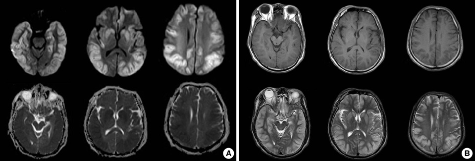
Fig. 1 The initial magnetic resonance imaging. (A) Diffusion-Weighted MRI of the brain showed multiple bilateral hyperintense signals along the cortical and subcortical regions (frontal, temporal, parietal, and occipital lobes), hippocampus, caudate, globus pallidus, and putament and ADC (afferent diffusion coefficient) map showed low signal at the same area. (B) Low signal intensity lesion at T1-weighted image and High signal intensity lesion at T2 weighted image were seen at same area (Fig. 1B).
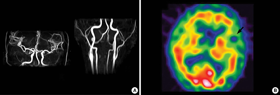
Fig. 2 Angiography and single photon emission computed tomography (SPECT). (A) MR angiography of the brain and neck showed no abnormalities. (B) The SPECT with 99mTc-HMPAO showed focal hypoperfusion in the left temporal lobe.

Fig. 3 The follow-up magnetic resonance imaging. (A) Follow-up images on the 20th day show revisal of the hyperintensity lesions on diffusion-MRI and hypointensity lesion on ADC map curvilinear. (B) The T1 and T2-weighted image showed linear high signal intensity selectively along the cortical regions of bilateral hemisphere and basal ganglia.
Reference
-
1. Cho SJ, Minn YK, Kwon KH. Severe hypoglycemia and vulnerability of the brain. Arch Neurol. 2006. 63:138.2. Aoki T, Sato T, Hasegawa K, Ishizaki R, Saiki M. Reversible hyperintensity lesion on diffusion-weighted MRI in hypoglycemic coma. Neurology. 2004. 63:392–393.3. Anderson JM, Milner RD, Strich SJ. Pathological changes in the nervous system in severe neonatal hypoglycaemia. Lancet. 1966. 2:372–375.4. Yoneda Y, Yamamoto S. Cerebral cortical laminar necrosis on diffusion-weighted MRI in hypoglycaemic encephalopathy. Diabet Med. 2005. 22:1098–1100.5. Chan R, Erbay S, Oljeski S, Thaler D, Bhadelia R. Case report: hypoglycemia and diffusion-weighted imaging. J Comput Assist Tomogr. 2003. 27:420–423.6. de Courten-Myers GM, Xi G, Hwang JH, Dunn RS, Mills AS, Holland SK, Wagner KR, Myers RE. Hypoglycemic brain injury: potentiation from respiratory depression and injury aggravation from hyperglycemic treatment overshoots. J Cereb Blood Flow Metab. 2000. 20:82–92.7. Christiaens FJ, Mewasingh LD, Christophe C, Goldman S, Dan B. Unilateral cortical necrosis following status epilepticus with hypoglycemia. Brain Dev. 2003. 25:107–112.8. Boeve BF, Bell DG, Noseworthy JH. Bilateral temporal lobe MRI changes in uncomplicated hypoglycemic coma. Can J Neurol Sci. 1995. 22:56–58.9. Fujioka M, Okuchi K, Hiramatsu KI, Sakaki T, Sakaguchi S, Ishii Y. Specific changes in human brain after hypoglycemic injury. Stroke. 1997. 28:584–587.10. Bakshi R, Morcos MF, Gabryel TF, Dandona P. Is fluid-attenuated inversion recovery MRI more sensitive than conventional MRI for hypoglycemic brain injury? Neurology. 2000. 55:1064–1065.11. Finelli PF. Diffusion-weighted MR in hypoglycemic coma. Neurology. 2001. 57:933.12. Spar JA, Lewine JD, Orrison WW Jr. Neonatal hypoglycemia: CT and MR findings. AJNR Am J Neuroradiol. 1994. 15:1477–1478.13. Barkovich AJ, Ali FA, Rowley HA, Bass N. Imaging patterns of neonatal hypoglycemia. AJNR Am J Neuroradiol. 1998. 19:523–528.14. Cakmakci H, Usal C, Karabay N, Kovanlikaya A. Transient neonatal hypoglycemia: cranial US and MRI findings. Eur Radiol. 2001. 11:2585–2588.15. Myers RE, Khan KJ. Bradley JB, Meldrum BS, editors. Insulin-induced hypoglycemia in the nonhuman primate. II: Long-term neuropathological consequences. Brain hypoxia (chapter 20). 1971. 39/40. 195–206. Reprinted in Clin Devel Med.16. Liwnicz BH, Mouradian MD, Ball JB Jr. Intense brain cortical enhancement on CT in laminar necrosis verified by biopsy. AJNR Am J Neuroradiol. 1987. 8:157–159.17. Tippin J, Adams HP Jr, Smoker WR. Early computed tomographic abnormalities following profound cerebral hypoxia. Arch Neurol. 1984. 41:1098–1100.18. Arbelaez A, Castillo M, Mukherji SK. Diffusion-weighted MR imaging of global cerebral anoxia. AJNR Am J Neuroradiol. 1999. 20:999–1007.19. Siskas N, Lefkopoulos A, Ioannidis I, Charitandi A, Dimitriadis A. Cortical laminar necrosis in brain infarcts: serial MRI. Neuroradiology. 2003. 45:283–288.20. Lovblad KO, Wetzel SG, Somon T, Wilhelm K, Mehdizade A, Kelekis A, El-Koussy M, El-Tatawy S, Bishof M, Schroth G, Perrig S, Lazeyras F, Sztajzel R, Terrier F, Rufenacht D, Delavelle J. Diffusion-weighted MRI in cortical ischaemia. Neuroradiology. 2004. 46:175–182.21. Iizuka T, Sakai F, Suzuki N, Hata T, Tsukahara S, Fukuda M, Takiyama Y. Neuronal hyperexcitability in stroke-like episodes of MELAS syndrome. Neurology. 2002. 59:816–824.22. Adams JH, Brierley JB, Connor RC, Treip CS. The effect of systemic hypotension upon the human brain. Clinical and neuropathological observation in 11 cases. Brain. 1966. 89:235–268.
- Full Text Links
-
- Actions
-
Cited
- CITED
-
- Close
- Share
-
- Similar articles
-
- Cortical Laminar Necrosis in an Infant with Severe Traumatic Brain Injury
- MRI Findings of Cortical Laminar Necrosis
- Acute Hepatic Encephalopathy Presenting as Cortical Laminar Necrosis: Case Report
- Cortical Laminar Necrosis associated with Osmotic Demyelination Syndrome
- Rapid Regression of White Matter Changes in Hypoglycemic Encephalopathy
