J Korean Soc Spine Surg.
2015 Dec;22(4):178-182. 10.4184/jkss.2015.22.4.178.
Motor Weakness of Right Ankle Dorsiflexion Caused by Increasing Size of Sacroiliac Joint Cyst after Posterior Lumbar Interbody Fusion in a Patient with Spinal Stenosis: A Case Report
- Affiliations
-
- 1Department of Orthopeadic Surgery, National Police Hospital, Seoul, Korea. osahnyj@lycos.co.kr
- KMID: 2150546
- DOI: http://doi.org/10.4184/jkss.2015.22.4.178
Abstract
- STUDY DESIGN: Case report
OBJECTIVES
To report a case of motor weakness caused by the increasing size of a sacroiliac joint cyst after spinal fusion. SUMMARY OF LITERATURE REVIEW: There have been no reports on the increased size of a sacroiliac joint cyst and motor weakness after spinal fusion.
MATERIALS AND METHODS
A 63-year-old female was admitted with low back pain and right sciatica. Magnetic resonance imaging (MRI) findings showed the spinal canal narrowing at L4-5 and a cystic lesion on the right sacroiliac joint. After surgery, the symptoms were relieved.
RESULTS
One month after the operation, motor function had worsened to grade 4. Follow-up MRI revealed an increase in the size of the cystic lesion. Selective nerve root blocks were performed. There was gradual improvement, and the motor grade reached grade 5 seven months after the operation.
CONCLUSIONS
We recommend that surgeons evaluate the adjacent segmental lesion by MRI before performing spinal fusion.
Keyword
MeSH Terms
Figure
-

Fig. 1. Initial magnetic resonance imaging (MRI) reveals spinal stenosis at L4–5 in the sagittal (A) and the axial (B) images, and a cystic lesion measuring 1.6 cm×1.0 cm at the inferior surface of the right sacroiliac joint in the coronal (C) image.

Fig. 2. Posterior decompression and posterior lumbar interbody fusion with a cage was performed at L4–5 (A, B).

Fig. 3. Followup MRI revealed the increased size of the cystic lesion measuring 3.3 cm×2.2 cm×1.6 cm in coronal (A) and axial (B) images (inferior and ventral aspects of the sacroiliac joint, S2–3 level).
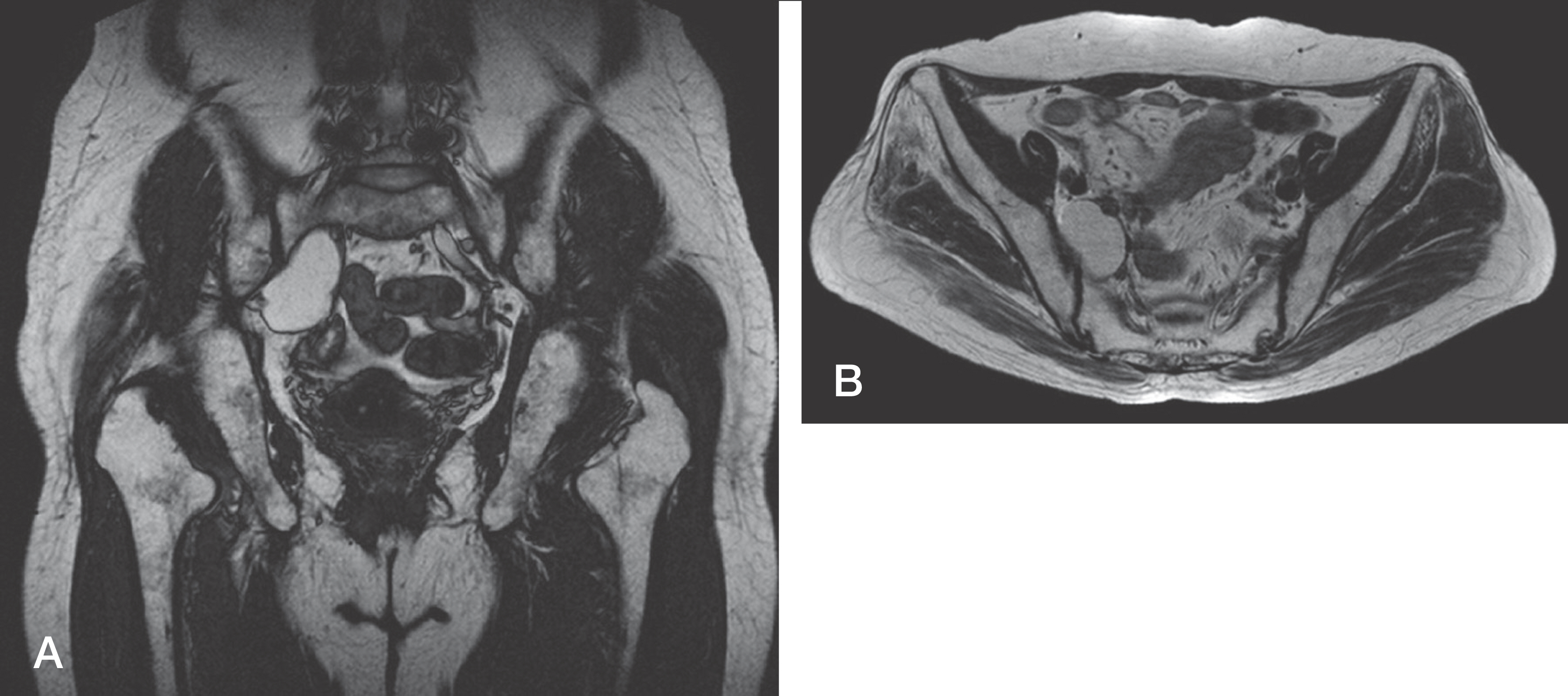
Fig. 4. Followup MRI taken after three years of the operation showed a further increase in the lesion size to 4.9 cm×2.6 cm×1.8 cm in the coronal (A) and axial (B) images.
Reference
-
1. Ha KY, Kim YH, Kang KS. Surgery for Adjacent Segment Changes after Lumbosacral Fusion. J Korean Soc Spine Surg. 2002; 9:332–40.
Article2. Hwang CJ, Lee SW, Ahn YJ, et al. Risk Factors for Adjacent Segment Disease After Lumbar Fusion. J Korean Soc Spine Surg. 2008; 15:44–53.
Article3. Bastian L, Lange U, Knop C, et al. Evaluation of The Mobility of Adjacent Segments after Posterior Thoracolumbar Fixation: A Biomechanical Study. Eur Spine J. 2001; 10:295–300.
Article4. Park P, Garton HJ, Gala VC, et al. Adjacent Segment Disease after Lumbar or Lumbosacral Fusion: Review of the Literature. Spine (Phila Pa 1976). 2004; 29:1938–44.
Article5. Ha KY, Lee JS, Kim KW. Degeneration of Sacroiliac Joint After Instrumented Lumbar or Lumbosacral Fusion: A Prospective Cohort Study over Five-Year Followup. Spine (Phila Pa 1976). 2008; 33:1192–8.6. Kikuchi S, Konno S, Kayama S, et al. Increased Resistance to Acute Compression Injury in Chronically Compressed Spinal Nerve Roots: An Experimental Study. Spine (Phila Pa 1976). 1996; 21:2544–50.
- Full Text Links
-
- Actions
-
Cited
- CITED
-
- Close
- Share
-
- Similar articles
-
- Clinical Comparison between Decompression and Posterior Lumbar Interbody Fusion in Chronic Lower Back Pain Involving Degenerative Disc Disease and Spinal Stenosis
- Minimally Invasive Lateral Lumbar Interbody Fusion: Indications, Outcomes and Complications
- Cauda Equina Syndrome Caused by Bilateral Facet Cyst Accompanying Spinal Stenosis
- Comparison between Posterior and Transforaminal Approaches for Lumbar Interbody Fusion
- Clinical Comparison between Microsurgical Decompression and Lumbar Interbody Fusion with Instrumentation for Lumbar Stenosis
