Anterior Abdominal Wall Leiomyoma Arising De Novo in a Fertile Women: A Case Report
- Affiliations
-
- 1Department of Radiology, Hallym University College of Medicine, Kangnam Sacred Heart Hospital, Seoul, Korea. baccas@hallym.or.kr
- 2Department of Pathology, Hallym University College of Medicine, Kangnam Sacred Heart Hospital, Seoul, Korea.
- KMID: 2150466
- DOI: http://doi.org/10.3348/jksr.2016.74.1.71
Abstract
- Abdominal wall leiomyoma arising de novo is very rare, hence the reported imaging findings of this disease are also rare. We reported the case of a 33-year-old woman who presented with an abdominal wall mass without antecedent gynecological surgeries. The initial abdominal computed tomography (CT) showed thickening of the left rectus abdominis and the loss of intervening fat between the rectus abdominis and the lateral abdominal muscles. After 8 months, the follow-up contrast-enhanced CT and ultrasonography (US) showed a lentiform-shaped mass with isodensity to the adjacent muscles. The US-guided biopsy was consistent with leiomyoma.
MeSH Terms
Figure
-
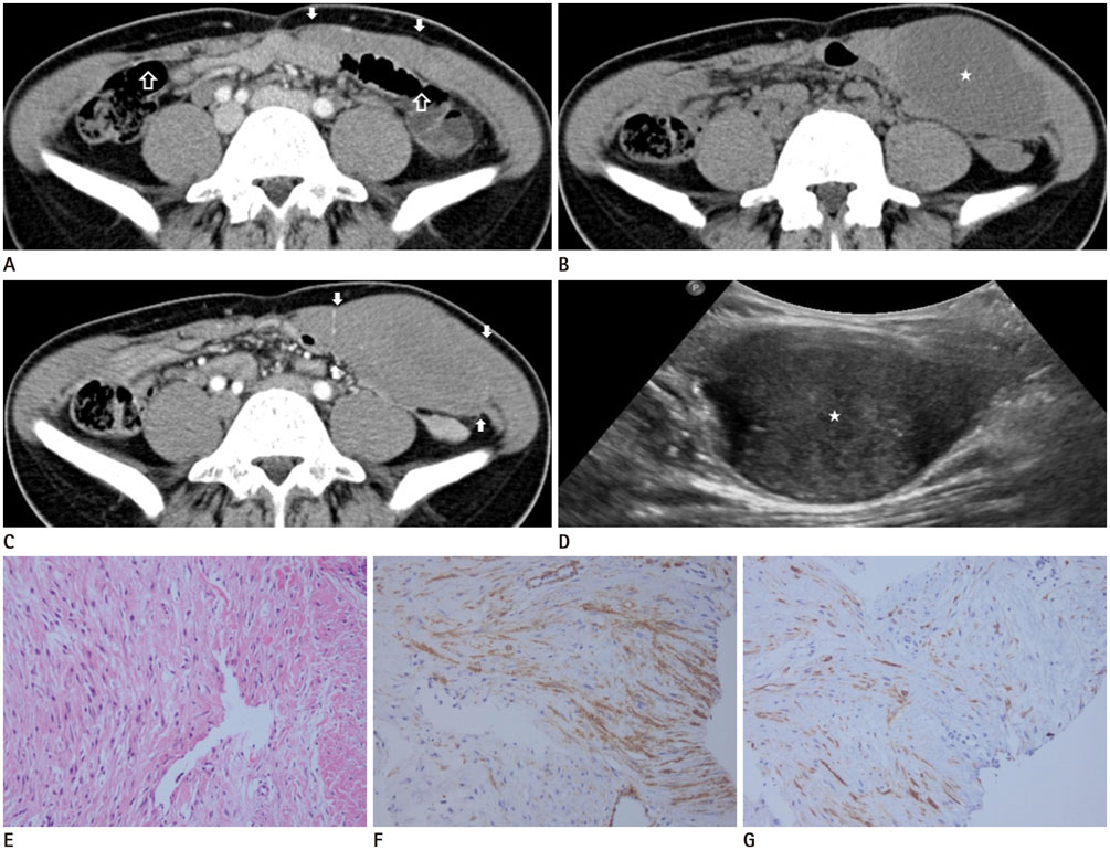
Fig. 1 A 34-year-old woman presenting with a palpable abdominal mass. A. The contrast-enhanced axial CT scan shows thickening of the left rectus abdominis muscle (arrows) and loss of intervening fat between the left rectus abdominis and the lateral abdominal wall muscles in contrast with the other side (open arrows). B. After 8 months, nonenhanced axial CT scan reveals a well-circumscribed mass (asterisk) with hypodensity, as compared with adjacent muscles. C. On the contrast-enhanced axial CT scan, the mass (arrows) is homogeneously enhanced and nearly isodense to the adjacent muscles. It is located in the left rectus abdominis and lateral abdominal muscles. D. The ultrasonography image shows a well-defined and mixed hypoechoic mass with a lentiform shape (asterisk). E. Microscopy of the needle biopsy specimen shows the diffuse proliferation of spindle-shaped cells but no atypia (hematoxylin and eosin, × 100). F, G. In the immunohistochemical stain, the spindle-shaped cells show immunoreactivity for smooth muscle actin and desmin (× 200).
Reference
-
1. Kjerulff KH, Langenberg P, Seidman JD, Stolley PD, Guzinski GM. Uterine leiomyomas. Racial differences in severity, symptoms and age at diagnosis. J Reprod Med. 1996; 41:483–490.2. Stewart EA. Uterine fibroids. Lancet. 2001; 357:293–298.3. Moon HS, Koo JS, Park SH, Park GS, Choi JG, Kim SG. Parasitic leiomyoma in the abdominal wall after laparoscopic myomectomy. Fertil Steril. 2008; 90:1201.e1–1201.e2.4. Ostrzenski A. Uterine leiomyoma particle growing in an abdominal-wall incision after laparoscopic retrieval. Obstet Gynecol. 1997; 89(5 Pt 2):853–854.5. Kumar S, Sharma JB, Verma D, Gupta P, Roy KK, Malhotra N. Disseminated peritoneal leiomyomatosis: an unusual complication of laparoscopic myomectomy. Arch Gynecol Obstet. 2008; 278:93–95.6. Ono M, Inoue Y, Yokota M, Uehara I, Kamijo S, Hattori Y, et al. Abdominal wall leiomyoma in a reproductive age woman without antecedent pelvic surgery. Eur J Obstet Gynecol Reprod Biol. 2010; 151:225–226.7. Midya M, Dewanda NK. Primary anterior abdominal wall leiomyoma-a diagnostic enigma. J Clin Diagn Res. 2014; 8:NJ01–NJ02.8. Al-Wadaani HA. Anterior abdominal wall leiomyoma arising de novo in a perimenopausal woman. Oman Med J. 2012; 27:323–325.9. Patterson GK, Winburn GB. Abdominal wall endometriomas: report of eight cases. Am Surg. 1999; 65:36–39.10. Einstein DM, Tagliabue JR, Desai RK. Abdominal desmoids: CT findings in 25 patients. AJR Am J Roentgenol. 1991; 157:275–279.
- Full Text Links
-
- Actions
-
Cited
- CITED
-
- Close
- Share
-
- Similar articles
-
- A Case of Leiomyoma of the Vagina
- Endometriosis and myoma concurrently arising after laparoscopic subtotal hysterectomy
- A Case of Leiomyoma of the Vagina
- A Case of Leiomyoma of the Vaginal Wall in a Proximal Suburethral and Subtrigonal Location Causing Voiding Difficulty
- A case of leiomyoma in the female urethra
