Clin Endosc.
2015 Sep;48(5):374-379. 10.5946/ce.2015.48.5.374.
Image Quality Analysis of Various Gastrointestinal Endoscopes: Why Image Quality Is a Prerequisite for Proper Diagnostic and Therapeutic Endoscopy
- Affiliations
-
- 1Digestive Disease Center, CHA Bundang Medical Center, CHA University, Seongnam, Korea. cjy6695@chamc.co.kr
- 2Digestive Disease Center, Soonchunhyang University Hospital, Soonchunhyang University College of Medicine, Seoul, Korea.
- KMID: 2148547
- DOI: http://doi.org/10.5946/ce.2015.48.5.374
Abstract
- Arising from human curiosity in terms of the desire to look within the human body, endoscopy has undergone significant advances in modern medicine. Direct visualization of the gastrointestinal (GI) tract by traditional endoscopy was first introduced over 50 years ago, after which fairly rapid advancement from rigid esophagogastric scopes to flexible scopes and high definition videoscopes has occurred. In an effort towards early detection of precancerous lesions in the GI tract, several high-technology imaging scopes have been developed, including narrow band imaging, autofocus imaging, magnified endoscopy, and confocal microendoscopy. However, these modern developments have resulted in fundamental imaging technology being skewed towards red-green-blue and this technology has obscured the advantages of other endoscope techniques. In this review article, we have described the importance of image quality analysis using a survey to consider the diversity of endoscope system selection in order to better achieve diagnostic and therapeutic goals. The ultimate aims can be achieved through the adoption of modern endoscopy systems that obtain high image quality.
Keyword
MeSH Terms
Figure
-
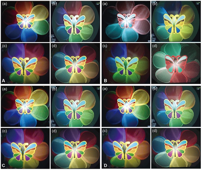
Fig. 1 Butterfly pattern (in vitro) taken according to each endoscopy company, analyzed on each point of evaluation, namely (A) definition, (B) boundary, (C) brightness, and (D) light and shade.

Fig. 2 Gastric lesion (in vivo) taken according to each endoscopy company analyzed on each point of evaluation, namely (A) definition, (B) boundary, (C) brightness, and (D) light and shade.
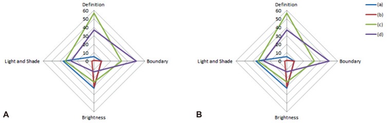
Fig. 3 Comparison of preference per product. (A) Butterfly pattern. (B) Gastric lesion.
Reference
-
1. Classen M, Knyrim K, Seidlitz HK, Hagenmüller F. Electronic endoscopy: the latest technology. Endoscopy. 1987; 19:118–123. PMID: 3608920.2. Bruno MJ. Magnification endoscopy, high resolution endoscopy, and chromoscopy: towards a better optical diagnosis. Gut. 2003; 52(Suppl 4):iv7–iv11. PMID: 12746262.
Article3. Muto M, Horimatsu T, Ezoe Y, Morita S, Miyamoto S. Improving visualization techniques by narrow band imaging and magnification endoscopy. J Gastroenterol Hepatol. 2009; 24:1333–1346. PMID: 19702901.
Article4. Tajiri H, Matsuda K, Fujisaki J. What can we see with the endoscope? Present status and future perspectives. Dig Endosc. 2002; 14:131–137.
Article5. Sano Y, Muto M, Tajiri H, Ohtsu A, Yoshida S. Optical/digital chromoendoscopy during colonoscopy using narrow-band imaging system. Dig Endosc. 2005; 17(Suppl 1):S43–S48.
Article6. Cho WY, Jang JY, Lee DH. Endoscopic Technology and Investigation Study Group. Recent advances in image-enhanced endoscopy. Clin Endosc. 2011; 44:65–75. PMID: 22741116.
Article7. Kodashima S, Fujishiro M. Novel image-enhanced endoscopy with i-scan technology. World J Gastroenterol. 2010; 16:1043–1049. PMID: 20205272.
Article8. Yamaguchi M, Haneishi H, Ohyama N. Beyond red-green-blue (rgb): spectrum-based color imaging technology. J Imaging Sci Technol. 2008; 52:10201-1–10201-15.9. Osawa H, Yoshizawa M, Yamamoto H, et al. Optimal band imaging system can facilitate detection of changes in depressed-type early gastric cancer. Gastrointest Endosc. 2008; 67:226–234. PMID: 18061596.
Article10. Yoshizawa M, Osawa H, Yamamoto H, et al. Diagnosis of elevated-type early gastric cancers by the optimal band imaging system. Gastrointest Endosc. 2009; 69:19–28. PMID: 19111685.
Article11. Demling L, Hagel HJ. Video endoscopy. Fundamentals and problems. Endoscopy. 1985; 17:167–169. PMID: 4054060.12. Yao K, Oishi T, Matsui T, Yao T, Iwashita A. Novel magnified endoscopic findings of microvascular architecture in intramucosal gastric cancer. Gastrointest Endosc. 2002; 56:279–284. PMID: 12145613.
Article
- Full Text Links
-
- Actions
-
Cited
- CITED
-
- Close
- Share
-
- Similar articles
-
- Recent Update of Gastrointestinal Endoscope Reprocessing
- Image quality and artifacts in automated breast ultrasonography
- The Past, Present, and Future of Image-Enhanced Endoscopy
- Introduction to Starting Upper Gastrointestinal Endoscopy: Proper Insertion, Complete Observation, and Appropriate Photographing
- Contrast Does Not Affect Cholangioscope Image Quality
