Diagnostic and Prognostic Implications of Spine Magnetic Resonance Imaging at Diagnosis in Patients with Multiple Myeloma
- Affiliations
-
- 1Department of Internal Medicine, Chungnam National University Hospital, Daejeon, Korea. deogyeon@cnu.ac.kr
- 2Department of Diagnostic Radiology, Chungnam National University Hospital, Daejeon, Korea.
- KMID: 2148496
- DOI: http://doi.org/10.4143/crt.2014.010
Abstract
- PURPOSE
The aim of this study is to determine the diagnostic and prognostic role of baseline spinal magnetic resonance imaging (MRI) in patients with multiple myeloma.
MATERIALS AND METHODS
We enrolled patients newly diagnosed with multiple myeloma from 2004-2011 at a single center. Abnormal MRI findings that were not detected in radiographs have been analyzed and categorized as malignant compression fractures or extramedullary plasmacytoma. The bone marrow (BM) infiltration patterns on MRI have been classified into five categories.
RESULTS
A total of 113 patients with a median age of 65 years (range, 40 to 89 years) were enrolled in the study. Malignant compression fractures not detected in the bone survey were found in 26 patients (23.0%), including three patients (2.6%) with no related symptoms or signs. Extramedullary plasmacytoma was detected in 22 patients (19.5%), including 15 (13.3%) with epidural extension of the tumor. Of these 22 patients, 11 (50.0%) had no relevant symptoms or signs. The presence of malignant compression fractures did not influence overall survival; whereas non-epidural extramedullary plasmacytoma was associated with poor overall survival in the multivariate analysis (hazard ratio, 3.205; 95% confidence interval [CI], 1.430 to 9.845; p=0.042). During the follow-up for a median of 21 months (range, 1 to 91 months), overall survival with the mixed BM infiltrative pattern (median, 24.0 months; 95% CI, 22.9 to 25.1 months) was shorter than those with other patterns (median 56 months; 95% CI, 48.9 to 63.1 months; p=0.030).
CONCLUSION
These results indicate that spine MRI at the time of diagnosis is useful for detecting skeletal lesions and predicting the prognosis in patients with multiple myeloma.
MeSH Terms
Figure
-

Fig. 1. (A) Malignant compression fracture, which was defined as a collapsed body with contour bulging of the involved masses. Extra-medullary extension of plasmacytoma, including epidural extension (B, indicated by an arrow) and non-epidural extension (C, indicated by arrows).
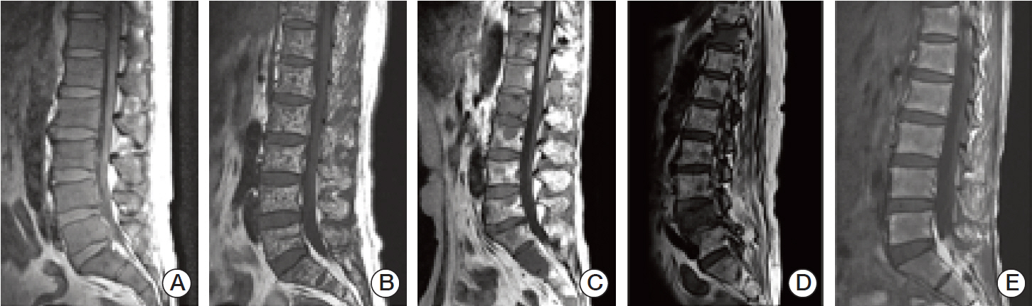
Fig. 2. (A) Diffuse infiltrative pattern. Homogeneous hypointense change which is similar or lower than disc signal on T1-weighted image. (B) Micronodular pattern, also referred to as “salt-and-pepper” pattern. Small foci of low intensity throughout the marrow on sagittal T1-weighted image. (C) Macronodular pattern. Multifocal relatively large hypointense lesions are detected on sagittal T1-weighted image. (D) Mixed pattern. It shows a combined micronodular and macronodular lesions. (E) Normal bone marrow pattern. Bone marrow shows hyperintense compared to the disc on T1-weighted image.
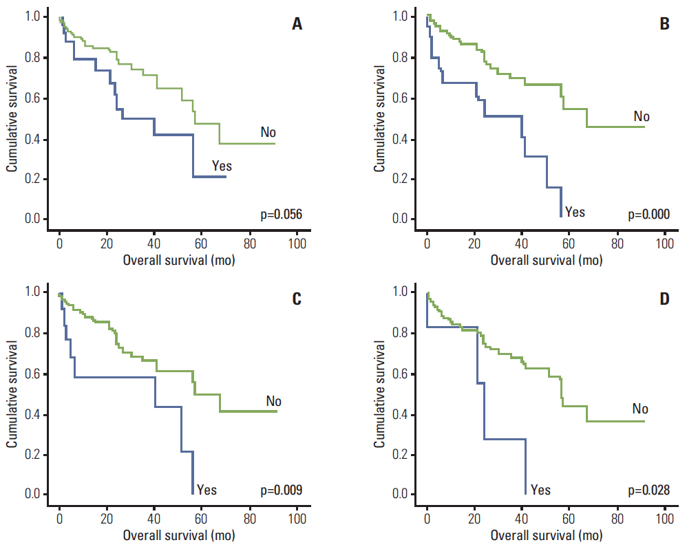
Fig. 3. Kaplan-Meier plots of survival according to magnetic resonance imaging abnormalities. (A) Survival according to malignant compression fracture. (B) Survival according to extramedullary extension of plasmacytoma. (C) Survival according to epidural extension of plasmacytoma. (D) Survival according to non-epidural extension of plasmacytoma.

Fig. 4. Kaplan-Meier plots of survival according to bone marrow infiltrative patterns. (A) Survival according to each of bone marrow infiltrative patterns. (B) Survival according to mixed patterns and others.
Cited by 1 articles
-
Clinical impact of spine magnetic resonance imaging as a valuable prognostic tool for patients with multiple myeloma: a retrospective study
Jung Min Lee, Hee Jeong Cho, Joon-Ho Moon, Sang Kyun Sohn, Byunggeon Park, Dong Won Baek
J Yeungnam Med Sci. 2022;39(4):300-308. doi: 10.12701/jyms.2021.01648.
Reference
-
References
1. Palumbo A, Anderson K. Multiple myeloma. N Engl J Med. 2011; 364:1046–60.
Article2. Bird JM, Owen RG, D'Sa S, Snowden JA, Pratt G, Ashcroft J, et al. Guidelines for the diagnosis and management of multiple myeloma 2011. Br J Haematol. 2011; 154:32–75.
Article3. Dimopoulos M, Kyle R, Fermand JP, Rajkumar SV, San Miguel J, Chanan-Khan A, et al. Consensus recommendations for standard investigative workup: report of the International Myeloma Workshop Consensus Panel 3. Blood. 2011; 117:4701–5.
Article4. D'Sa S, Abildgaard N, Tighe J, Shaw P, Hall-Craggs M. Guidelines for the use of imaging in the management of myeloma. Br J Haematol. 2007; 137:49–63.5. Baur-Melnyk A, Buhmann S, Durr HR, Reiser M. Role of MRI for the diagnosis and prognosis of multiple myeloma. Eur J Radiol. 2005; 55:56–63.
Article6. Moulopoulos LA, Gika D, Anagnostopoulos A, Delasalle K, Weber D, Alexanian R, et al. Prognostic significance of magnetic resonance imaging of bone marrow in previously untreated patients with multiple myeloma. Ann Oncol. 2005; 16:1824–8.
Article7. Dimopoulos M, Terpos E, Comenzo RL, Tosi P, Beksac M, Sezer O, et al. International myeloma working group consensus statement and guidelines regarding the current role of imaging techniques in the diagnosis and monitoring of multiple Myeloma. Leukemia. 2009; 23:1545–56.
Article8. Hillengass J, Landgren O. Challenges and opportunities of novel imaging techniques in monoclonal plasma cell disorders: imaging "early myeloma". Leuk Lymphoma. 2013; 54:1355–63.
Article9. Tan E, Weiss BM, Mena E, Korde N, Choyke PL, Landgren O. Current and future imaging modalities for multiple myeloma and its precursor states. Leuk Lymphoma. 2011; 52:1630–40.
Article10. Baur-Melnyk A, Buhmann S, Becker C, Schoenberg SO, Lang N, Bartl R, et al. Whole-body MRI versus whole-body MDCT for staging of multiple myeloma. AJR Am J Roentgenol. 2008; 190:1097–104.
Article11. Zamagni E, Nanni C, Patriarca F, Englaro E, Castellucci P, Geatti O, et al. A prospective comparison of 18F-fluorodeoxyglucose positron emission tomography-computed tomography, magnetic resonance imaging and whole-body planar radiographs in the assessment of bone disease in newly diagnosed multiple myeloma. Haematologica. 2007; 92:50–5.
Article12. Fonti R, Salvatore B, Quarantelli M, Sirignano C, Segreto S, Petruzziello F, et al. 18F-FDG PET/CT, 99mTc-MIBI, and MRI in evaluation of patients with multiple myeloma. J Nucl Med. 2008; 49:195–200.
Article13. Walker R, Barlogie B, Haessler J, Tricot G, Anaissie E, Shaughnessy JD Jr, et al. Magnetic resonance imaging in multiple myeloma: diagnostic and clinical implications. J Clin Oncol. 2007; 25:1121–8.
Article14. Hanrahan CJ, Christensen CR, Crim JR. Current concepts in the evaluation of multiple myeloma with MR imaging and FDG PET/CT. Radiographics. 2010; 30:127–42.
Article15. Stabler A, Baur A, Bartl R, Munker R, Lamerz R, Reiser MF. Contrast enhancement and quantitative signal analysis in MR imaging of multiple myeloma: assessment of focal and diffuse growth patterns in marrow correlated with biopsies and survival rates. AJR Am J Roentgenol. 1996; 167:1029–36.16. Kusumoto S, Jinnai I, Itoh K, Kawai N, Sakata T, Matsuda A, et al. Magnetic resonance imaging patterns in patients with multiple myeloma. Br J Haematol. 1997; 99:649–55.
Article17. Baur A, Stabler A, Nagel D, Lamerz R, Bartl R, Hiller E, et al. Magnetic resonance imaging as a supplement for the clinical staging system of Durie and Salmon? Cancer. 2002; 95:1334–45.
Article18. Hillengass J, Wasser K, Delorme S, Kiessling F, Zechmann C, Benner A, et al. Lumbar bone marrow microcirculation measurements from dynamic contrast-enhanced magnetic resonance imaging is a predictor of event-free survival in progressive multiple myeloma. Clin Cancer Res. 2007; 13(2 Pt 1):475–81.19. Moulopoulos LA, Dimopoulos MA, Alexanian R, Leeds NE, Libshitz HI. Multiple myeloma: MR patterns of response to treatment. Radiology. 1994; 193:441–6.
- Full Text Links
-
- Actions
-
Cited
- CITED
-
- Close
- Share
-
- Similar articles
-
- Diffusion-Weighted Magnetic Resonance Imaging of Spine
- A Case of Nonsecretory Multiple Myeloma with Atypical Imaging Features
- Analysis of Bone Mineral Density in Multiple Myeloma: A Comparison of Bone Mineral Density with Plain Radiography, Magnetic Resonance Imaging, and Clinical Staging
- Nonsecretory Multiple Myeloma with Multiple Spine Fracture
- Clinical impact of spine magnetic resonance imaging as a valuable prognostic tool for patients with multiple myeloma: a retrospective study
